Understanding Follicular Miniaturisation: Causes, Signs, and Science
9 min read ·
Understanding Follicular Miniaturisation explains how hair follicles shrink over time, why DHT drives the process, and what evidence-based steps halt pattern thinning.
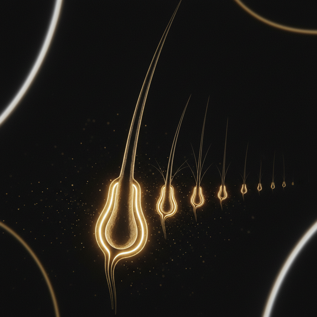
Quick answer: Understanding Follicular Miniaturisation is essential for recognising pattern hair loss, as it describes the biological process where hair follicles progressively shrink over repeated growth cycles. Driven primarily by dihydrotestosterone (DHT) in genetically susceptible individuals, affected follicles produce increasingly thinner, shorter, and less pigmented hairs until they may stop producing visible fibres entirely. The main limitation is that once a follicle undergoes complete fibrosis or senescent atrophy, the miniaturisation process becomes irreversible by standard non-surgical means.
Key takeaways
- Follicular miniaturisation is the hallmark histological feature of androgenetic alopecia in both men and women.
- It transforms terminal hairs (thick, deeply pigmented fibres) into vellus-like hairs (fine, short, unpigmented strands).
- The androgen dihydrotestosterone (DHT), synthesised by the enzyme 5-alpha reductase, acts as the chief trigger in genetically predisposed follicles.
- Miniaturisation is distinct from diffuse telogen shedding; it involves a progressive reduction in follicular diameter and growth phase duration, not sudden shedding.
- Early clinical intervention can halt or partially reverse miniaturisation, but completely fibrosed follicles cannot regrow terminal fibres.
Understanding Follicular Miniaturisation: what the evidence shows
Understanding Follicular Miniaturisation begins with examining how healthy hair follicles behave over successive cycles. Under normal physiological conditions, a terminal scalp hair follicle cycles through anagen (active growth, lasting 2 to 7 years), catagen (regression, lasting 2 to 3 weeks), and telogen (resting, lasting 2 to 4 months). When a new cycle begins, the dermal papilla reconstructs a matrix capable of producing a hair shaft with a typical diameter of 60 to 90 micrometres.
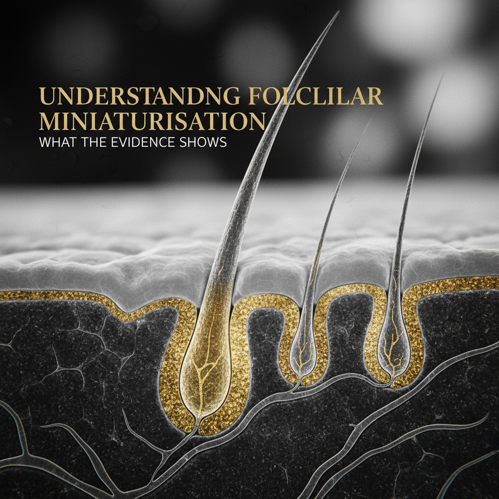
In androgenetic alopecia, this regenerative process deteriorates. In genetically susceptible scalp regions, such as the frontal hairline, temples, and crown, dihydrotestosterone (DHT) binds to androgen receptors within the cells of the dermal papilla. This binding alters gene expression and upregulates inhibitory signalling cascades, most notably transforming growth factor-beta (TGF-beta) and Dickkopf-related protein 1 (DKK-1).
These signalling factors prematurely truncate the anagen phase and lengthen the kenogen phase (the lag phase between telogen shedding and the emergence of a new anagen fibre). With each consecutive, shortened cycle, the volume of the dermal papilla shrinks. Because dermal papilla cell number directly determines hair shaft calibre, the resulting hair fibres become progressively thinner, reaching diameters under 30 micrometres. Over years, terminal hairs transform into wispy, vellus-like hairs.
The biological mechanisms behind follicle shrinking
The physiological shift from a robust terminal follicle to a miniaturised unit involves several concurrent biological events:
- Enzymatic conversion of testosterone: Local follicular tissues express 5-alpha reductase (primarily Type II). This enzyme reduces testosterone into dihydrotestosterone (DHT), an androgen with approximately five times greater affinity for the androgen receptor.
- Dermal papilla signal alteration: Once DHT occupies androgen receptors in the dermal papilla, paracrine signalling shifts. Inhibitory proteins suppress the Wnt/beta-catenin pathway, which is essential for maintaining follicular stem cell proliferation and anagen duration.
- Shortened anagen duration: Instead of remaining in growth for several years, affected hairs may exit anagen after only several months or weeks. Consequently, the hair shaft never achieves its genetic potential for length or thickness.
- Perifollicular microinflammation: Biopsies of miniaturising scalp tissue frequently reveal low-grade, perifollicular lymphocytic infiltrate around the follicular infundibulum. This microinflammation can accelerate tissue remodelling and connective tissue sheath thickening.
- Eventual follicular fibrosis: If the process continues unchecked for years, the dermal sheath collapses and the follicular tract is replaced by non-functional fibrous connective tissue (a follicular scar or stela). At this stage, stem cells in the hair follicle bulge can no longer interact with an intact dermal papilla.
Terminal hairs vs vellus-like hairs
Miniaturisation creates distinct structural shifts in the hair fibre that can be observed both clinically and under magnification:

| Characteristic | Healthy Terminal Hair | Miniaturised / Vellus-Like Hair |
|---|---|---|
| Shaft Diameter | 60 to 90 micrometres | Less than 30 micrometres |
| Anagen Duration | 2 to 7 years | Few weeks to several months |
| Internal Medulla | Present and continuous/fragmented | Completely absent |
| Pigmentation | Fully pigmented by melanocytes | Pale, reduced, or unpigmented |
| Follicular Depth | Extends 4 to 5 mm into subcutis | Sits high in reticular dermis (1 to 2 mm) |
| Arrector Pili Muscle | Firmly attached to follicle bulge | Loose, altered, or detached connection |
Clinical signs: how to identify miniaturisation
Miniaturisation rarely happens overnight. Because it affects neighbouring follicles asynchronously, the scalp initially displays a wide variation in hair shaft diameters across a single region.
- Hair diameter diversity (anisotrichosis): When viewing the scalp with a trichoscope, anisotrichosis is the earliest diagnostic sign of miniaturisation. If more than 20 percent of hairs in an affected area show varying diameters, androgenetic alopecia is typically present.
- Single-hair follicular units: Healthy scalps feature follicular units containing groupings of two, three, or four hairs. As individual follicles within a unit miniaturise and stop growing, units gradually diminish to single-hair groupings.
- Yellow dots: Trichoscopy may show follicular openings filled with sebum and keratin debris where a hair has failed to regrow, indicating empty or severely miniaturised follicles.
- Decreased scalp coverage without shedding: Individuals often notice that the scalp becomes visible under bright light or when wet, even though they have not observed large clumps of hair falling out in the shower or brush.
What the evidence does not show
Popular discussions around follicle shrinking frequently lead to misconceptions about its causes and progression. Controlled research clarifies several crucial boundaries:
- It is not caused by poor surface circulation alone: While microvascular changes occur in miniaturised zones, they are secondary consequences of follicular atrophy, not the primary initiating cause. Simply stimulating blood flow without addressing androgenic signalling does not reverse the condition.
- It is not normal shedding: Telogen effluvium involves a temporary synchronisation of hairs into the shedding phase due to physiological stress, nutritional deficits, or illness. Telogen effluvium produces high shed counts of full-thickness terminal hairs. Miniaturisation, by contrast, is a structural reduction of the follicle itself.
- Shampoo and sebum do not cause miniaturisation: Excess sebum and scalp oils do not choke follicles or trigger genetic shrinking. While sebum contains small amounts of DHT, follicular miniaturisation is driven by intracellular androgen receptors and local enzymatic activity deep within the follicle bulb.
Management options: supporting follicle health
Managing miniaturisation relies on early action. Scientific evidence confirms that halting follicular shrinking is considerably easier than regenerating an already miniaturised follicle.
Medical therapies
Prescription and evidence-based treatments target the biological mechanisms directly:
- 5-alpha reductase inhibitors: Oral or topical formulations reduce systemic or scalp DHT levels, decreasing androgen receptor activation in the dermal papilla.
- Potassium channel openers (Minoxidil): Topical and low-dose oral minoxidil shorten the telogen phase and prolong anagen, widening follicular diameters and extending growth periods.
- Low-level light therapy (LLLT): Photobiomodulation using specific red light wavelengths (650 to 670 nanometres) stimulates mitochondrial activity and adenosine triphosphate (ATP) production in dermal papilla cells.
Scalp care and cosmetic maintenance
Topical products cannot replace pharmacological treatments for severe genetic miniaturisation, but maintaining an optimal follicular environment supports fibre resilience:
- Scalp hygiene: Routine washing with gentle, non-stripping cleansers removes inflammatory build-up, oxidised sebum, and debris that can exacerbate perifollicular stress.
- Topical botanicals: Caffeine, rosemary extract, niacinamide, and biotin are common additions to scalp routines. Controlled lab studies indicate caffeine can counteract certain DHT-mediated suppression in vitro, although cosmetic scalp tonics serve primarily to support a healthier scalp feel and improve hair manageability.
Watermans scalp support
For those seeking to maintain a healthy scalp environment while addressing thinning or fragile hair, Watermans offers the Grow More Elixir and Grow Me Shampoo. Formulated with targeted cosmetic ingredients including biotin, caffeine, rosemary extract, niacinamide, and lupin protein, these formulations are designed to nourish the scalp surface, strengthen existing hair fibres against mechanical breakage, and promote a visibly thicker, fuller appearance. They serve as supportive cosmetic options alongside broader trichological care.
When hair loss is caused by something else
Not all thinning involves androgen-driven miniaturisation. It is critical to differentiate between conditions that require entirely different management strategies:
- Telogen effluvium: Marked by abrupt, widespread shedding across the entire scalp, typically emerging 2 to 3 months after a trigger such as severe illness, surgery, childbirth, or rapid weight loss. Hair diameters remain uniform.
- Alopecia areata: An autoimmune disorder where T-cells attack anagen follicles, causing sudden, well-defined circular patches of complete hair loss. Exclamation mark hairs, rather than miniaturised fibres, appear at the margins.
- Cicatricial (scarring) alopecias: Conditions such as lichen planopilaris or frontal fibrosing alopecia involve irreversible inflammatory destruction of the follicular stem cells. They show scalp erythema, perifollicular scale, and a complete absence of follicular ostia, requiring immediate medical intervention to prevent scarring.
- Traction alopecia: Caused by prolonged, repetitive mechanical tension from tight hairstyles. It primarily affects the frontal and temporal margins and leads to follicular trauma rather than hormone-driven shrinking.
When to seek professional advice
You should consult a GP, registered trichologist, or dermatologist when:
- Thinning progresses rapidly over a period of weeks or months.
- You observe scalp symptoms such as persistent redness, itching, burning, flaking, or pain.
- Hair loss appears in distinct, patchy circles or presents alongside eyebrow or eyelash loss.
- You want an accurate trichoscopic evaluation to measure follicular diameter diversity and establish a clinical treatment plan before permanent fibrosis develops.
Frequently asked questions about Understanding Follicular Miniaturisation
Can a miniaturised hair follicle be fully restored?
If a follicle has only partially miniaturised, medical therapies such as minoxidil or 5-alpha reductase inhibitors can often halt the process and partially increase shaft diameter and anagen length. However, once a follicle has undergone complete miniaturisation into a secondary vellus state or transitioned into connective tissue fibrosis, it can no longer be restored to a full terminal hair.
How does Understanding Follicular Miniaturisation help in diagnosing male vs female pattern hair loss?
Understanding Follicular Miniaturisation clarifies why patterns differ between sexes. In men, miniaturisation typically concentrates along the hairline and vertex due to high localized receptor density, often leading to total baldness in those zones. In women, miniaturisation is usually diffuse across the crown and central parting with preservation of the frontal hairline, rarely progressing to complete bareness.
Does frequent hair washing worsen follicular miniaturisation?
No. Frequent washing does not accelerate miniaturisation. Washing merely dislodges hairs that have already detached from the follicular matrix and are sitting in the telogen phase. Follicular miniaturisation occurs deep within the dermis and is driven by genetics and hormones, not by water, shampoo, or the physical action of cleansing the scalp.
What is the difference between hair shedding and hair miniaturisation?
Hair shedding refers to hair fibres completing their cycle and falling from the scalp, typically 50 to 100 hairs per day. Miniaturisation is a biological structural change where the follicle itself shrinks over years. A person undergoing miniaturisation may experience little to no abnormal daily shedding, yet their overall hair volume gradually decreases because each replacement hair is finer and shorter.
Summary
Understanding Follicular Miniaturisation allows both professionals and individuals to look beyond superficial hair care and address the cellular mechanisms of pattern hair loss. By recognising that progressive thinning stems from androgen-induced cycle shortening and dermal papilla regression, you can seek timely clinical diagnostics and implement targeted therapies before follicles reach irreversible fibrosis.
Sources and references
- British Association of Dermatologists. (2020). Androgenetic Alopecia: Clinical Guideline for Management.
- National Institute for Health and Care Excellence (NICE). (2023). Alopecia, androgenetic - Clinical Knowledge Summaries.
- Journal of the American Academy of Dermatology. (2017). Trichoscopy of androgenetic alopecia: A systematic review. DOI: 10.1016/j.jaad.2017.02.045.
- Dermatology Practical & Conceptual. (2021). Anisotrichosis and miniaturization: Pathognomonic signs in hair loss diagnosis. DOI: 10.5826/dpc.1102a42.
- International Journal of Molecular Sciences. (2020). The Role of Androgens and Androgen Receptors in Follicular Miniaturization. DOI: 10.3390/ijms21124317.
Find these ingredients in Watermans products
- Niacinamide: you will find this ingredient in our Hair Mask.