Trichorrhexis Nodosa Overview: Causes, Signs, and Management
10 min read ·
A comprehensive Trichorrhexis Nodosa Overview detailing the causes, paintbrush-like microscopic signs, mechanical mechanisms, and evidence-based repair strategies.
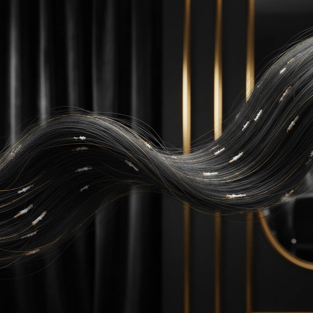
Quick answer: A comprehensive Trichorrhexis Nodosa Overview reveals that this condition is the most common structural hair shaft defect, characterised by fragile swelling nodes where the cuticle fails and cortical fibres splay apart like two interlocking paintbrushes. This defect causes mechanical breakage along the fibre rather than true follicular hair loss. The main limitation is that split cortical fibres cannot biologically re-fuse; resolving the issue requires identifying environmental or metabolic triggers, halting cumulative physical and chemical trauma, and allowing healthy new growth to replace damaged lengths.
Key takeaways
- Structural defect, not follicular loss: Trichorrhexis nodosa represents physical hair breakage along the shaft, leaving the living hair follicle entirely intact.
- Microscopic paintbrush appearance: Under dermoscopy or light microscopy, the characteristic nodes resemble two frayed brushes thrust into each other.
- Acquired trauma is primary: Most cases stem from cumulative mechanical, thermal, and chemical stress, including excessive heat styling, bleaching, and vigorous brushing.
- Congenital forms exist: Rare inherited disorders, such as Netherton syndrome, trichothiodystrophy, and argininosuccinic aciduria, present with congenital hair fragility.
- Cuticle loss triggers the node: Stripping the protective outer lipid and cuticular layers exposes cortical keratin bundles, which splay outward under standard tensile tension.
- Irreversible fibre damage: Once a fibre develops nodes, that specific segment cannot be permanently repaired; management relies on protective grooming, conditioning, and trims.
Trichorrhexis Nodosa Overview: what the evidence shows
In clinical trichology, an accurate Trichorrhexis Nodosa Overview begins with the architecture of the human hair fibre. Hair consists of three concentric zones: the outermost cuticle, the structural cortex, and the central medulla. The cortex provides tensile strength through parallel keratin intermediate filaments embedded in a high-sulphur matrix.
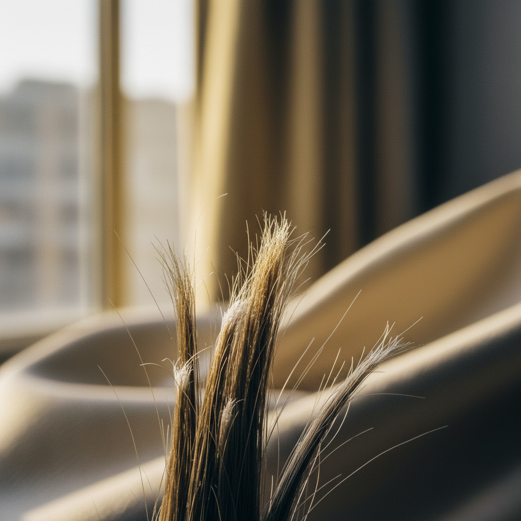
Research published across clinical dermatology confirms that trichorrhexis nodosa develops when physical, chemical, or systemic factors disrupt the cuticle. When the cuticle wears away, the exposed cortical cells lose their cohesion. Under routine physical forces such as combing, drying, or resting on a pillow, the longitudinal cortical fibrils splay laterally. This creates tiny, bead-like nodules visible along the shaft. When mechanical tension exceeds the reduced tensile strength of these exposed fibres, the hair snaps cleanly at the node.
Clinicians distinguish between three presentation patterns:
- Proximal trichorrhexis nodosa: Seen frequently in Afro-textured hair, where anatomical curvature, low moisture content, and chemical relaxers lead to breakage close to the scalp (often within 1 to 5 centimetres).
- Distal trichorrhexis nodosa: Common in straight and wavy hair types, presenting as tiny white specks and severe splitting at the outer ends of long hair due to years of cumulative weathering, ultraviolet exposure, and heat styling.
- Congenital or generalised trichorrhexis nodosa: Appears in early infancy or childhood, often accompanied by sparse growth, developmental delay, or metabolic abnormalities.
| Feature | Trichorrhexis Nodosa | Monilethrix | Trichorrhexis Invaginata | Normal Weathering |
|---|---|---|---|---|
| Microscopic Appearance | Frayed paintbrushes colliding | Beaded variations in shaft diameter | "Bamboo hair" (ball-and-socket invagination) | Jagged fracture, mild cuticle lifting |
| Primary Location | Mid-shaft or distal tips | Diffuse across scalp | Diffuse, eyebrows often involved | Distal 5 to 10 cm of hair ends |
| Typical Cause | Physical/chemical trauma; rarely metabolic | Genetic (KRT81, KRT83, KRT86 mutations) | Genetic (SPINK5 mutation / Netherton syndrome) | Environmental wear, friction |
| Follicular Health | Normal follicles, intact density | Follicular keratosis, perifollicular erythema | Follicular plugging, atopic skin | Completely normal follicles |
| Reversibility | Resolves once trauma ceases and hair regrows | Chronic, genetic lifelong course | Chronic, managed symptomatically | Trim damaged ends, apply conditioning |
The biological mechanism of node formation
The structural breakdown in trichorrhexis nodosa follows a clear biomechanical progression. Healthy hair fibres rely on 18-methyleicosanoic acid (18-MEA), a protective lipid covalently bonded to the outer epicuticle. This lipid layer gives the cuticle a hydrophobic surface with a natural contact angle above 100 degrees, reducing friction between adjacent strands.
When hair undergoes repeated alkaline treatments (such as bleaching at pH 9.0 to 10.5 or chemical straightening at pH 12.0 to 14.0), the 18-MEA barrier is stripped. The hair fibre becomes hydrophilic, absorbing water rapidly and swelling. Repeated cycles of swelling and drying cause hygral stress, lifting the cuticular scales.
Once cuticular scale margins break off, mechanical friction directly impacts the cortex. The cortex relies on disulphide bonds formed by cystine residues for its integrity. Thermal styling tools operating at temperatures above 185 degrees Celsius cause structural changes to cortical alpha-keratin, turning it into disordered beta-sheet keratin. Stripped of its cuticle and weakened by heat, the parallel bundle of macrofibrils buckles under everyday tension. The fibres splay outwards, creating the macroscopic white nodule.
What the evidence does not show
It is critical to clarify what scientific literature does not support regarding this condition.
First, the evidence does not show that trichorrhexis nodosa is an inflammatory follicular disease. It does not cause scarring of the scalp, it does not destroy follicular stem cells, and it does not halt anagen (growth) phase activity. Telogen counts remain normal unless an unrelated telogen effluvium coexists.
Second, topical cosmetic products cannot permanently weld ruptured cortical fibrils back together. While conditioning agents, hydrolysed proteins, and film-forming polymers coat the shaft and temporarily reduce friction, they do not reverse internal mechanical fractures. Claims that any cosmetic wash can "cure" structural nodes contradict basic hair biology.
Third, trichorrhexis nodosa is not caused by poor hygiene, microbial scalp infections, or routine shampooing with balanced formulations. Washing hair cleanly does not cause cortical collapse unless accompanied by aggressive towel scrubbing, boiling water, or harsh detangling.
Signs and clinical presentation to look for
Identifying trichorrhexis nodosa requires observing both visual cues and tactile feedback:
- White specks along the hair: Patients and stylists often observe tiny, pale nodes along the shaft. These are frequently mistaken for head lice nits, trichobacteriosis, or dandruff flakes. Unlike nits, which adhere to one side of the shaft, a node encompasses the entire circumference.
- Short, uneven lengths: The hair appears patchy or seems to have stopped growing. In reality, hair continues to emerge from the follicle at the normal rate (roughly 1 to 1.25 centimetres per month), but fractures before reaching length.
- Increased shedding of short fragments: Sufferers notice small, blunt-ended hair fragments on pillows, clothing, and sinks, differing from natural telogen shed hairs that feature an intact white bulb at the root.
- Dull, coarse texture: The hair feels dry, tangles easily, and lacks natural light reflection due to widespread cuticle degradation.
Managing and resolving the condition
Because the hair fibre is non-living tissue, damaged segments cannot biologically heal. Management requires protecting existing strands and allowing unaffected new growth to replace fractured lengths.
1. Eliminating physical and thermal friction
Cease all direct heat styling above 150 degrees Celsius. Let hair dry naturally where practical, or diffuse on a low-heat setting. Replace stiff bristle brushes with wide-tooth, seamless combs, detangling only when hair is coated with a conditioning lubricant. Avoid vigorous towel drying; blot gently with a microfibre cloth instead.
2. Adjusting chemical processes
Discontinue chemical relaxers, perms, and oxidative bleaching while the condition is active. If colouring is essential, switch to deposit-only, non-ammonia demi-permanent dyes that deposit pigment without lifting the cuticle at high alkaline levels.
3. Restoring surface lubrication and pH balance
Use conditioning agents formulated between pH 4.5 and 5.5 to smooth remaining cuticular margins. Cationic conditioning agents (such as cetrimonium chloride and behentrimonium methosulphate) deposit positively charged complexes onto negatively charged damaged sites, temporarily reducing surface friction by up to 50 percent. Leave-in silicone polymers or plant-derived emollients provide a protective sacrificial barrier against grooming friction.
4. Strategic trimming
Cutting the hair shaft just above the highest visible node eliminates the point of mechanical weakness, preventing fractures from propagating further down the fibre.
When the cause may be an underlying metabolic issue
While the vast majority of trichorrhexis nodosa presentations are acquired through grooming trauma, sudden fragility without a history of physical abuse warrants medical investigation.
Congenital metabolic defects can disrupt keratinisation:
- Argininosuccinic aciduria: A defect in the urea cycle that starves keratin of arginine, causing severe hair fragility, developmental delays, and elevated blood ammonia.
- Menkes kinky hair disease: An X-linked copper transport deficiency that impairs lysyl oxidase, an enzyme essential for keratin cross-linking.
- Hypothyroidism and iron deficiency: Systemic hormone or ferritin deficits compromise hair fibre calibre and quality, rendering emerging hair far more vulnerable to minor daily grooming friction.
When to seek professional advice
Consult a dermatologist, trichologist, or general practitioner if:
- Hair snaps continuously despite eliminating heat, chemicals, and mechanical stress for 8 to 12 weeks.
- White nodes appear across infant or early-childhood hair.
- Breakage is accompanied by systemic signs such as fatigue, cold intolerance, unexplained weight shifts, brittle nails, or intellectual delay.
- The scalp exhibits erythema, scaling, pustules, or localised bald patches.
A specialist will perform light microscopy, dermoscopy, or polarised light examination on plucked and clipped strands to verify whether nodes reflect trichorrhexis nodosa, monilethrix, or another structural anomaly.
Support from Watermans cosmetic haircare
For individuals recovering from acquired mechanical hair breakage, supporting the scalp environment while maintaining cosmetic condition helps new hair emerge smoothly. Watermans formulations focus on scalp comfort and cosmetic fibre vitality. Products like Watermans Grow Me shampoo and Condition Me conditioner provide a balanced, gentle wash free from aggressive sulphates, delivering hydrolysed lupin protein, niacinamide, and caffeine to condition the hair surface, reduce combing resistance, and keep new growth feeling robust as damaged ends are steadily trimmed away.
Frequently asked questions about Trichorrhexis Nodosa Overview
Can a hair mask repair hair affected by trichorrhexis nodosa?
No. An evidence-based Trichorrhexis Nodosa Overview makes clear that once internal cortical fibres splay apart, no cosmetic mask, oil, or protein treatment can permanently fuse them back together. Conditioners and masks can coat the shaft, smooth cuticular borders, and reduce friction to shield undamaged segments from further stress, but fractured nodes must eventually be cut away.
Does trichorrhexis nodosa lead to permanent baldness?
No. Trichorrhexis nodosa affects the hair shaft, not the hair bulb or follicle buried beneath the scalp. Because the follicular stem cells remain healthy and undisturbed, hair continues to grow from the root. The apparent baldness or thinning seen in severe cases is an illusion caused by rapid fibre snapping close to the scalp surface.
How is trichorrhexis nodosa differentiated from head lice nits?
Under close inspection or dermoscopy, a trichorrhexis nodosa node forms a microscopic swelling that surrounds the entire diameter of the hair shaft. In contrast, a head lice nit is an oval casing glued firmly to one single side of the hair shaft at an acute angle. Nits also slip along the fibre when pulled, whereas nodes cannot be moved.
How long does it take for hair to recover from trichorrhexis nodosa?
Recovery time depends on how fast your hair grows and the length of your style. Hair typically grows at a rate of 1 to 1.25 centimetres per month. Once you eliminate aggressive heat, bleach, and mechanical trauma, new growth will emerge healthy. Complete recovery generally takes between 6 and 18 months as trimmed ends gradually replace the broken lengths.
Is trichorrhexis nodosa hereditary?
Most cases are acquired through routine environmental, chemical, and physical trauma. However, hereditary forms exist. Rare genetic disorders such as argininosuccinic aciduria, Netherton syndrome, and Menkes syndrome can produce congenital trichorrhexis nodosa. If hair fragility begins in early infancy alongside systemic symptoms, a medical genetics assessment is recommended.
Summary
In this Trichorrhexis Nodosa Overview, we have detailed the structural mechanism that causes cortical fibres to splay and break under everyday tension. This common defect is an acquired, external structural failure rather than an internal follicular disease. By setting aside damaging hot tools, avoiding aggressive chemical processes, adopting gentle detangling habits, and scheduling regular trims, you allow healthy, unbroken lengths to take the place of fragile strands.
Sources and references
- American Academy of Dermatology. Hair breakage and structural shaft abnormalities. AAD clinical resources.
- British Association of Dermatologists. Guidelines for the management of hair shaft defects.
- Dawber, R. (1996). Hair and Scalp Disorders: Common Presenting Signs, Differential Diagnosis and Management. Dermatologic Clinics.
- Whiting, D. A. (2004). Structural abnormalities of the hair shaft. Journal of the American Academy of Dermatology.
- National Institutes of Health (NIH). Genetic and Rare Diseases Information Center: Argininosuccinic Aciduria and Hair Manifestations.
Find these ingredients in Watermans products
- Copper: you will find this ingredient in our Hair Vitamins.