Scalp Biopsy Procedures: Diagnostic Steps, Histology, and Care
9 min read ·
Clinical Scalp Biopsy Procedures provide definitive diagnostic insight for complex hair loss. Learn how punch biopsies, histology, and recovery work.
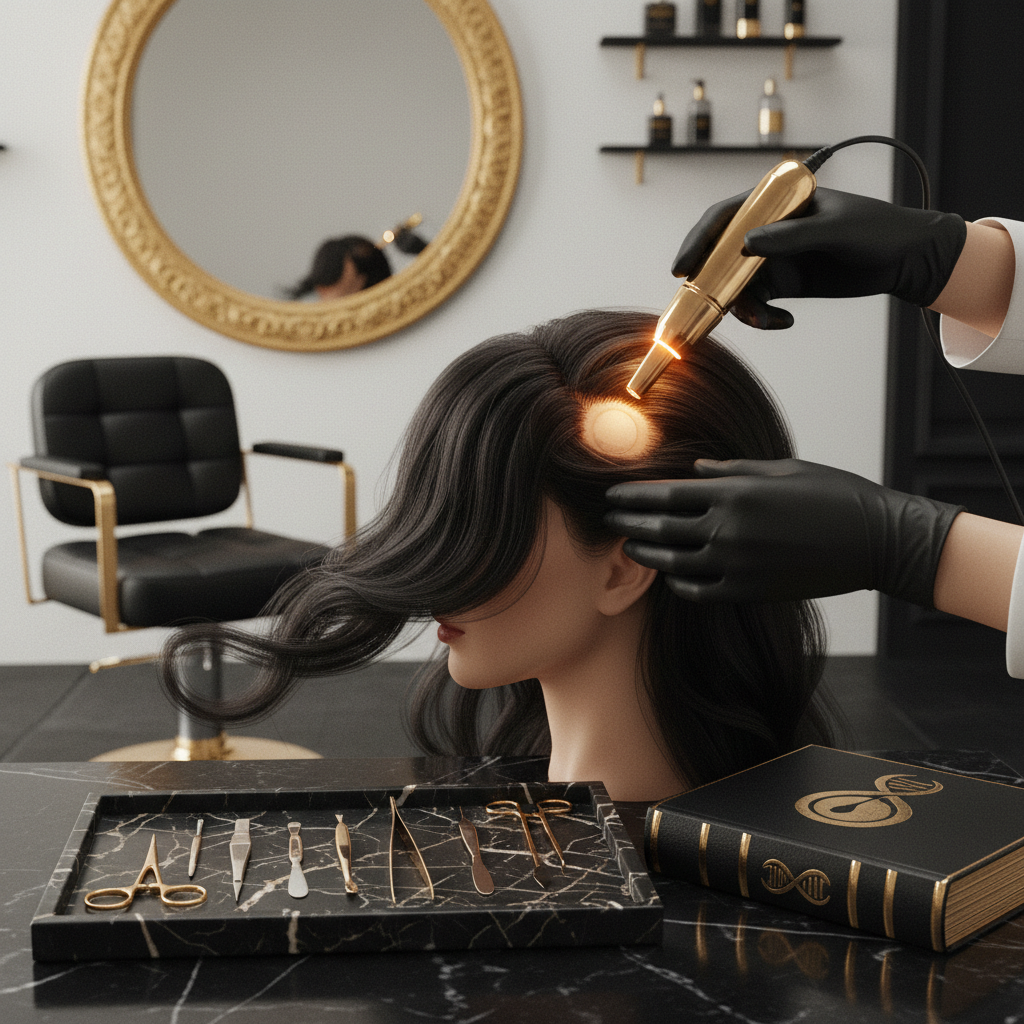
Quick answer: A scalp biopsy is a minor diagnostic surgical technique where a small sample of skin and hair follicles is removed under local anaesthetic for laboratory evaluation. Clinical Scalp Biopsy Procedures are the definitive gold standard for diagnosing complex alopecias, distinguishing scarring from non-scarring hair loss, and identifying inflammatory scalp disorders. The main limitation is that a biopsy provides a static snapshot of dynamic follicular pathology, meaning diagnostic accuracy relies entirely on precise site selection, proper mechanical sectioning, and skilled dermatopathological interpretation.
Key takeaways
- Scalp biopsies provide microscopic confirmation when clinical exams, trichoscopy, and blood tests cannot distinguish between overlapping hair loss disorders.
- The 4 mm punch biopsy is the clinical standard, penetrating full-thickness dermis down to the subcutaneous fat layer where terminal bulbs sit.
- Dual biopsies or transected specimens analysed via horizontal (transverse) and vertical (longitudinal) sectioning yield the highest diagnostic value.
- Differentiating cicatricial (scarring) alopecia from non-scarring alopecia is the most critical indication, as early diagnosis prevents irreversible follicular destruction.
- Recovery requires minimal downtime, with sutures generally removed within 7 to 14 days and negligible scarring when placed parallel to hair emergence angles.
Scalp Biopsy Procedures: what the evidence shows
Clinical Scalp Biopsy Procedures serve as the cornerstone of complex trichological diagnosis. Peer-reviewed literature from the British Association of Dermatologists and the American Academy of Dermatology consistently demonstrates that while non-invasive tools such as dermoscopy and trichoscopy guide clinical suspicion, histopathology remains necessary to resolve ambiguous presentations.
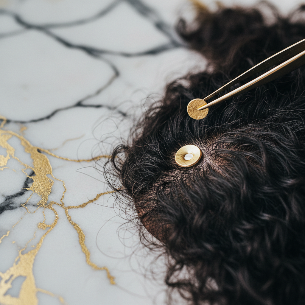
During the procedure, a clinician cleans the site with an antiseptic and infiltrates the dermis with local anaesthetic (typically 1% or 2% lidocaine with adrenaline to control bleeding). A circular punch blade, usually 4 mm in diameter, is rotated along the exit angle of the hair shaft. Aligning the blade precisely with follicular orientation is critical; inserting the punch at an incorrect angle transects follicular units midway down the shaft, destroying the diagnostic architecture needed to evaluate anagen-to-telogen ratios.
The core specimen must extend into the subcutaneous fat layer because terminal anagen hair bulbs reside 3 to 5 mm deep. Once excised, the wound is closed with one or two non-absorbable sutures or surgical staples. The tissue is then fixed in 10% neutral buffered formalin. To maximise diagnostic yield, modern dermatopathology relies on the Headington method of transverse (horizontal) sectioning, which allows total counts of terminal versus vellus follicles, alongside vertical sectioning to examine the dermo-epidermal junction and interfollicular epidermis.
What the evidence does not show
A biopsy is a powerful diagnostic tool, but published research highlights clear boundaries regarding what it can achieve:
- Predicting treatment speed or absolute regrowth: Histology confirms the current pathological state (such as follicular fibrosis, miniaturisation, or lymphocytic infiltrate). It cannot predict with absolute certainty whether dormant follicles will respond to medical therapies.
- Diagnosing diffuse shedding in isolation without clinical context: Telogen effluvium shows subtle histological shifts, such as an increase in the telogen count above 15% to 20%, without pathognomonic markers. A biopsy cannot pinpoint the systemic trigger (such as iron deficiency or acute illness) behind the shedding.
- Replacing routine clinical screening: Research does not support using biopsies as a first-line screen for straightforward male or female pattern hair loss. It is reserved for rapidly progressive, atypical, painful, or refractory cases.
The diagnostic mechanism: horizontal versus vertical sectioning
Understanding how pathologists examine tissue explains why technique matters so much. A standard 4 mm punch biopsy captures approximately 20 to 35 hair follicles, depending on donor density.
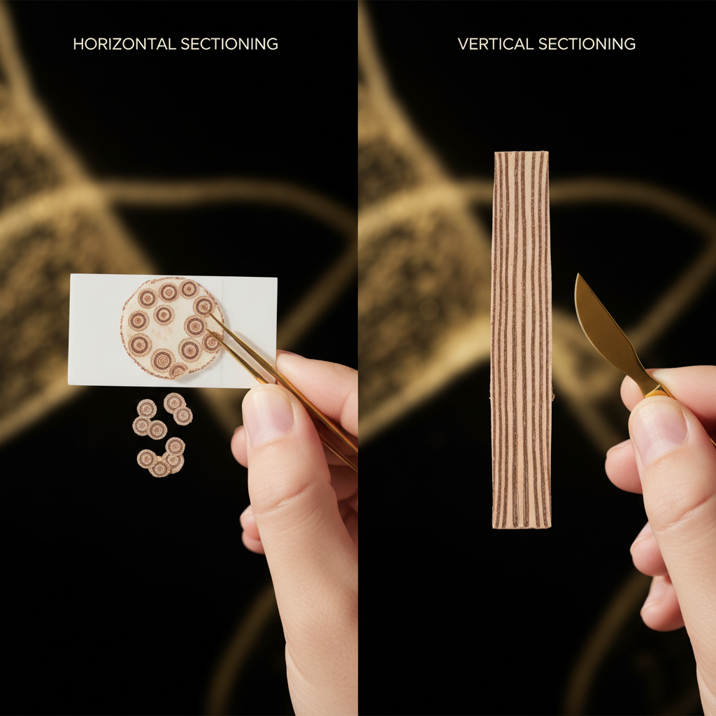
Vertical sections cut the tissue like a slice of bread from epidermis down to fat. This view is ideal for assessing epidermal changes, interface dermatitis, and structural shifts at the dermo-epidermal junction, common in conditions like discoid lupus erythematosus (DLE). However, a single vertical cut only captures a fraction of the follicles present in the tissue core.
Horizontal sectioning, introduced into routine practice by Dr Dawn Headington in 1984, cuts across the shafts parallel to the scalp surface at distinct anatomical levels: the infundibulum, isthmus, and subcutaneous fat. This view permits:
- Accurate calculation of the anagen-to-telogen ratio (normal scalp holds roughly 85% to 90% anagen follicles and under 10% to 15% telogen).
- Calculation of the terminal-to-vellus ratio (normally greater than 4:1, but dropping below 2:1 or even 1:1 in androgenetic alopecia).
- Assessment of fibrous tracts (streamers) indicating vanished follicles in severe alopecia areata or late-stage pattern hair loss.
| Feature | Horizontal (Transverse) Sectioning | Vertical (Longitudinal) Sectioning |
|---|---|---|
| Primary Strength | Quantitative analysis (total follicle counts, terminal-to-vellus ratios) | Architectural appraisal of epidermis and dermo-epidermal junction |
| Best For | Androgenetic alopecia, telogen effluvium, alopecia areata | Lichen planopilaris, discoid lupus, folliculitis decalvans |
| Follicular Yield | Evaluates virtually all follicles within the 4 mm core | Evaluates only the few follicles aligned with the blade plane |
| Depth Assessment | Stratified at infundibular, isthmic, and subcutaneous levels | Continuous vertical view from stratum corneum to adipose tissue |
| Clinical Standard | Often preferred for non-scarring hair loss | Required for inflammatory scarring alopcias |
When a biopsy is clinically indicated
Dermatologists do not perform biopsies on every patient presenting with hair thinning. The procedure is indicated when clinical examination leaves diagnostic uncertainty, particularly where prompt treatment is needed to prevent permanent hair loss:
- Suspected cicatricial (scarring) alopecia: Lichen planopilaris (LPP), frontal fibrosing alopecia (FFA), central centrifugal cicatricial alopecia (CCCA), or discoid lupus erythematosus (DLE). These conditions permanently destroy follicular stem cells at the bulge region.
- Unclear diffuse hair thinning: Distinguishing between diffuse alopecia areata, chronic telogen effluvium, and female pattern hair loss (FPHL).
- Unresponsive scalp conditions: Chronic scalp dermatoses that do not respond to standard anti-inflammatory or antifungal interventions.
- Presence of inflammatory signs: Perifollicular erythema, scaling, pustules, scalp induration, burning sensations, or loss of follicular ostia (open pores).
Choosing the biopsy site
Diagnostic accuracy depends heavily on site selection. For scarring alopcias, taking tissue from the completely bald, sclerotic centre of a patch is unhelpful; the lab will simply report non-specific end-stage fibrosis. Clinicians target the active advancing margin where early inflammation surrounds surviving follicles.
For non-scarring, diffuse disorders, clinicians take tissue from the area of greatest symptomatic involvement (often the vertex or crown), occasionally pairing it with a second control specimen from the clinically unaffected occipital scalp to establish the patient's individual baseline density.
When the cause may be something else
Hair shedding and thinning often stem from non-dermatological or purely mechanical causes that do not require invasive biopsy. Before undergoing minor surgery, ensure these possibilities have been explored:
- Hair breakage from mechanical or chemical stress: Breakage involves hair fibres fracturing along the shaft due to heat, bleach, or tension, rather than follicle-level dysfunction. The root remains securely in the scalp.
- Nutritional and endocrine shifts: Ferritin levels below 30 to 50 ug/L, thyroid dysfunction (elevated or suppressed TSH), and recent high fevers can provoke widespread shedding (telogen effluvium) without skin pathology.
- Traction alopecia: Persistent pulling forces on the hair follicle can cause hair loss without initial intrinsic follicular disease, identifiable early via patient styling history.
What to do: post-procedure care and recovery
Healing from a punch biopsy is straightforward. You will leave the clinic with a small dressing covering the site. Keep the area clean and dry for the first 24 to 48 hours. After this window, gentle cleansing with warm water and a mild, non-irritating cleanser is typically permitted, avoiding harsh scrubbing over the sutures.
Sutures are removed between day 7 and day 14. Bruising and mild swelling are normal; paracetamol is usually sufficient to manage minor discomfort. Avoid rigorous exercise, heavy sweating, hair dyes, and chemical straightening until the wound is fully closed and the sutures are removed.
Professional scalp support and cosmetic maintenance
While diagnostic Scalp Biopsy Procedures must be carried out by qualified dermatologists or surgeons, ongoing scalp care plays an important supportive role once healing is complete. Maintaining a clean, non-occlusive, balanced follicular environment supports hair fibres in remaining pliable and resilient.
For everyday cosmetic care, Watermans products provide gentle cleansing and conditioning designed to maintain a healthy-looking scalp and improve hair manageability. Formulated with revitalising botanical extracts, biotin, and hydrolysed proteins, Watermans shampoos and conditioners help reduce surface breakage and support fuller-looking hair without disrupting the delicate balance of the scalp barrier.
Frequently asked questions about Scalp Biopsy Procedures
Are Scalp Biopsy Procedures painful?
The procedure itself is largely painless because the clinician numbs the scalp with local anaesthetic before the punch tool is used. You will feel a brief stinging or pinching sensation during the local anaesthetic injection, which lasts roughly 10 to 20 seconds. Once numb, you might feel pressure or tugging, but no sharp discomfort. Mild soreness for a day or two afterwards is easily managed with simple over-the-counter pain relief.
Will a scalp biopsy leave a permanent scar?
Yes, any full-thickness incision through the dermis produces a tiny, permanent scar. However, because a 4 mm punch is small and typically closed with fine sutures parallel to the direction of surrounding hair growth, the resulting scar is usually minor and hidden by surrounding hair. Only in areas of extensive, complete baldness might the small pale dot remain visible upon close inspection.
How long does it take to get scalp biopsy results?
Results generally take between 1 and 3 weeks. After the tissue is taken, it must be fixed in formalin, embedded in paraffin wax, cut into micrometre-thin slices, and stained (often with haematoxylin and eosin, alongside special stains for elastic fibres or fungi). A specialised dermatopathologist must then examine and report on the slides.
Can a scalp biopsy diagnose telogen effluvium?
Yes, but it is rarely the first choice for diagnosis. A horizontal biopsy can confirm telogen effluvium by showing a shift where more than 15% to 20% of follicles are in the resting telogen phase without the significant miniaturisation seen in pattern hair loss. However, dermatologists prefer non-invasive tools such as pull tests, trichoscopy, and blood work before considering surgical intervention for diffuse shedding.
What should I avoid after having a scalp biopsy?
Avoid strenuous exercise, swimming, saunas, and washing the wound for the first 24 to 48 hours to minimise bleeding and infection risks. Do not apply harsh chemical hair dyes, perms, or heat styling tools near the biopsy site until the sutures have been removed and your clinician confirms the incision has fully healed.
Conclusion
When hair loss or scalp inflammation defies standard clinical evaluation, clinical Scalp Biopsy Procedures offer unmatched diagnostic clarity. By isolating a microscopic cross-section of hair follicles, these minor surgical investigations permit direct analysis of inflammatory patterns, follicular counts, and structural changes. Obtaining an accurate histological diagnosis gives your medical team the precise roadmap needed to preserve follicular health and protect your hair over the long term.
Sources and references
- British Association of Dermatologists. Clinical Guidelines for the Management of Alopecia Areata and Cicatricial Alopecias.
- Headington, J. T. (1984). Transverse microscopic anatomy of the human scalp: A basis for a morphometric approach to disorders of the hair follicle. Archives of Dermatology, 120(4), 449-456.
- American Academy of Dermatology Association. Diagnosing and Treating Hair Loss: Dermatopathologic Evaluation of Scalp Biopsies.
- National Institute for Health and Care Excellence (NICE). Alopecia: Diagnosis and Assessment in Primary and Secondary Care.
- Whiting, D. A. (1993). Diagnostic problems in alopecia: A clinician's perspective on the role of scalp biopsy. Dermatologic Clinics, 11(1), 35-46.
Find these ingredients in Watermans products
- Biotin: you will find this ingredient in our Hair Growth Shampoo.