Seborrhoeic Dermatitis on Deep Skin Tones: Symptoms, Mechanisms, and Safe Scalp Care
9 min read ·
Seborrhoeic Dermatitis on Deep Skin Tones often shows as hypopigmented or violaceous patches rather than red flaking. Learn the clinical signs, mechanisms, and care strategies.
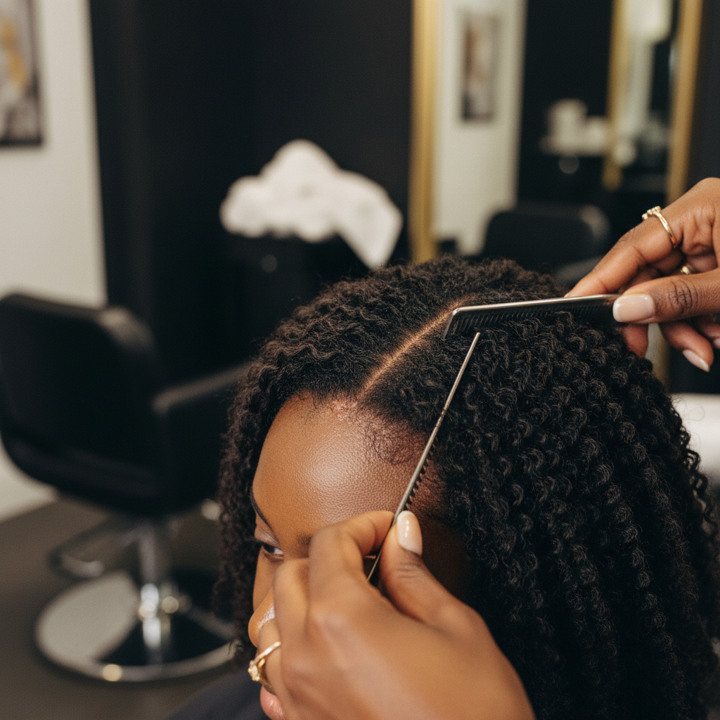
Quick answer: Seborrhoeic Dermatitis on Deep Skin Tones presents uniquely compared to lighter skin, frequently showing hypopigmentation (lightened patches), hyperpigmentation, or subtle violaceous plaques rather than classic bright erythema (redness). Managing Seborrhoeic Dermatitis on Deep Skin Tones requires balancing antifungal and anti-inflammatory therapy with the preservation of tight-textured hair moisture, as daily washing with drying medicated shampoos can cause severe shaft breakage. While medical treatments control active flares effectively, the main limitation is that the underlying tendency remains chronic and pigmentary changes can take months to resolve.
Key takeaways
- Atypical visual presentation: Erythema is often muted, dark brown, or violaceous on brown and Black skin, frequently accompanied by marked hypopigmentation that is mistaken for fungal infections like tinea versicolor.
- Hair texture considerations: Tightly coiled (Afro-textured) hair produces natural bends that impede sebum distribution, making the hair shafts prone to dry breakage if aggressive daily clarifying shampoos are used.
- Topical vehicle choice is critical: Alcohol-based scalp solutions and foams dry out tight curls, whereas cream-based, lotion, or targeted nozzle applications protect fibre hydration.
- Post-inflammatory pigment shifts: Scale removal must be gentle; aggressive scratching or peeling triggers post-inflammatory hyperpigmentation or hypopigmentation that outlasts the inflammation itself.
- Differentiating mimickers: Conditions such as tinea capitis, scalp psoriasis, discoid lupus erythematosus, and central centrifugal cicatricial alopecia (CCCA) must be carefully ruled out by a medical professional.
Seborrhoeic Dermatitis on Deep Skin Tones: what the evidence shows
Clinical dermatology literature indicates that Seborrhoeic Dermatitis on Deep Skin Tones often manifests with distinctive morphology that goes unrecognised in traditional textbook teaching. In Fitzpatrick skin types IV to VI, the inflammatory cascade triggered by Malassezia yeast does not produce the vivid, bright salmon-pink or red patches seen on fair skin. Instead, dermatologists observe dull pink, slate grey, violaceous, or hyperpigmented (darkened) plaques covered with greasy, yellowish or white scale.

Equally common on deep skin is petaloid seborrhoeic dermatitis. This variant forms delicate, polycyclic, ring-shaped patches that frequently appear hypopigmented (lighter than the surrounding skin). The hypopigmentation occurs because inflammatory mediators temporarily suppress melanocyte activity and transfer of melanosomes to surrounding keratinocytes, without permanently destroying the pigment cells.
Textbook clinical trials have historically tested daily ketoconazole or zinc pyrithione shampoos on straight, sebum-rich hair profiles. Peer-reviewed studies in ethnic dermatology demonstrate that applying this wash frequency to Afro-textured hair leads to severe fibre dehydration, cuticle chipping, and hair breakage along the shaft. Evidence confirms that clinical success in deeper skin phototypes relies on tailoring the vehicle, reducing wash frequency to once or twice weekly, and applying leave-on prescription agents directly to the skin rather than the hair fibres.
The underlying mechanism: Malassezia, sebum, and melanocytes
Seborrhoeic dermatitis is not caused by poor hygiene, nor is it a contagious infection. The condition develops from an abnormal inflammatory response to Malassezia yeasts (chiefly Malassezia globosa and Malassezia restricta), which are normal commensal micro-organisms living on human skin.
These lipid-dependent yeasts consume triglycerides within human sebum. Using lipase enzymes, Malassezia breaks triglycerides down into free fatty acids, notably oleic acid and arachidonic acid. In susceptible individuals, these unsaturated fatty acids penetrate the stratum corneum, compromising skin barrier integrity. The immune system mounts an inflammatory response, leading to rapid, disordered epidermal turnover, cellular parakeratosis (nucleated cells reaching the surface), and noticeable flaking.
On deep skin tones, this inflammatory cascade interacts directly with epidermal melanocytes:
- Inflammatory suppression: Inflammatory cytokines (such as IL-1 alpha and TNF-alpha) can inhibit tyrosinase activity, leading to post-inflammatory hypopigmentation.
- Melanocyte stimulation: In other presentations, intense inflammation and mechanical scratching activate melanocytes, depositing excess melanin into the dermis (pigment incontinence), resulting in persistent post-inflammatory hyperpigmentation.
- Barrier disruption: Coiled hair follicles naturally have a curved path through the dermis, and tight curl patterns make sebum migration along the hair shaft inefficient. This leaves the scalp oily while the hair fibre remains dry.
Clinical signs: identifying the condition on darker skin
Recognising the signs requires looking beyond standard textbook definitions of redness. On deep complexions, look for the following:
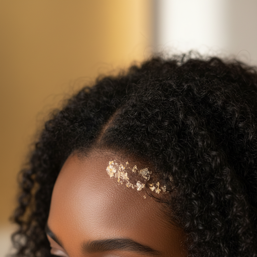
- Violaceous or slate-grey plaques: Discolouration along the anterior hairline, behind the ears, within the eyebrows, or across the nasolabial folds that looks purplish, dark brown, or grey rather than red.
- Macular hypopigmentation: Lightened patches around the hairline (the "seborrhoeic crown") or central scalp, often displaying fine, powdery, or greasy scaling.
- Petaloid configurations: Flower-petal-shaped, annular plaques across the face, chest, or scalp margins.
- Pruritus (itching): Burning or itching sensations that prompt scratching, which increases the risk of lichenification (skin thickening) and secondary bacterial infection.
- Scale characteristics: Flakes may range from fine and white to thick, adherent, and yellow-brown.
What the evidence does not show
Controlled research does not show that Malassezia colonisation levels are higher in individuals with deep skin tones compared to fair skin tones. The microbial density is comparable; what differs is the cutaneous immune reaction, the visibility of vascular erythema, and the melanocytic response to local inflammation.
Furthermore, evidence does not show that heavy hair greases, mineral oils, or raw plant butters cure seborrhoeic dermatitis. While traditional haircare practices for tight curls frequently involve "greasing the scalp" to soothe dryness, laboratory studies demonstrate that Malassezia yeasts can metabolise specific plant-derived fatty acids, potentially fuelling the flare. Applying heavy occlusive pomades can worsen follicular occlusion without addressing the underlying yeast or inflammation.
Finally, medical evidence does not support the idea that seborrhoeic dermatitis itself causes permanent, scarring hair loss. The condition affects the superficial epidermis. While severe itching and vigorous scratching can cause temporary hair shedding (telogen effluvium) or mechanical traction breakage, follicular loss only occurs if a misdiagnosed cicatricial alopecia is left untreated.
| Diagnostic & Care Factor | Presentation on Fair Skin (Types I, III) | Presentation on Deep Skin (Types IV, VI) |
|---|---|---|
| Primary colour of lesions | Bright pink, salmon-red, or scarlet erythema | Slate grey, dark brown, violaceous, or hypopigmented |
| Pigmentary aftermath | Transient erythema; rare long-term colour shift | Marked post-inflammatory hypo- or hyperpigmentation |
| Common pattern variant | Diffuse flaky patches across the scalp vertex | Petaloid (annular, flower-like) and hairline lesions |
| Wash tolerance | Tolerates daily washing with clarifying shampoos | Requires low-frequency washing (e.g. 1 to 2 times weekly) |
| Preferred formulation | Foams, solutions, gels, light shampoos | Creams, lotions, scalp-directed nozzle suspensions |
| Primary mimicker | Scalp psoriasis, contact dermatitis | Tinea capitis, discoid lupus, CCCA |
Practical management: balancing scalp care and hair fibre health
Managing Seborrhoeic Dermatitis on Deep Skin Tones demands a dual strategy: calming epidermal inflammation and yeast overgrowth while preventing hair fibre desiccation and breakage.
1. Tailored cleansing frequency
Standard guidance recommending daily medicated washing is damaging for Afro-textured and very curly hair. Wash the scalp once every 7 to 14 days, or at most twice weekly during acute flares. When washing, apply medicated cleansers (containing ketoconazole 2%, ciclopirox olamine 1%, zinc pyrithione 1%, or selenium sulfide) directly to the scalp skin using fingertips or an applicator bottle. Allow it to sit for 5 to 10 minutes so the active ingredient can penetrate the stratum corneum before rinsing thoroughly. Keep the lather off the lengths of the hair as much as possible.
2. Strategic vehicle selection
Request creams, lotions, or targeted oils from your prescribing physician rather than alcohol-based solutions, sprays, or light foams. Alcohol bases evaporate quickly but extract moisture from curly hair shafts, causing brittleness. Emulsion-based topical corticosteroids (such as fluocinolone acetonide scalp oil or mometasone furoate lotion) allow targeted anti-inflammatory action while respecting hair structure.
3. Careful moisturisation
Avoid thick, petrolatum-based pomades directly on the scalp skin during active flares, as they trap scales and can aggravate the microbial imbalance. Instead, apply nourishing leave-in conditioners and natural lipid sealants strictly to the mid-lengths and ends of the hair fibres, keeping the scalp clear.
4. Gentle descaling
Never scrape crusts off the scalp with fine-toothed combs, fingernails, or harsh bristles. Mechanical trauma induces the Koebner phenomenon (worsening skin lesions at sites of injury) and stimulates severe post-inflammatory hyperpigmentation. To soften thick scale, apply mineral oil or a designated scalp-softening formulation containing low-strength salicylic acid (1 to 2%), leave it on for 30 minutes under a warm towel, and rinse gently.
When the cause may be something else
Because Seborrhoeic Dermatitis on Deep Skin Tones presents with muted erythema and variable pigmentation, misdiagnosis is common. Several conditions must be considered:
- Tinea capitis: A true dermatophyte fungal infection that is common in children and can occur in adults. Unlike seborrhoeic dermatitis, tinea capitis frequently causes patchy hair breakage near the scalp surface ("black dot" sign), swollen cervical lymph nodes, and intense local inflammation. A fungal culture or scalp scraping distinguishes the two.
- Central Centrifugal Cicatricial Alopecia (CCCA): A progressive scarring alopecia seen almost exclusively in women of African descent. It begins at the crown or vertex and spreads outward, accompanied by burning, tenderness, or itching. It permanently destroys follicles. If flaking is accompanied by hair thinning and scalp shine, an immediate dermatology evaluation with a scalp biopsy is essential.
- Discoid Lupus Erythematosus (DLE): This autoimmune condition produces violaceous, indurated plaques that heal with central depigmentation, scarring, follicular plugging, and permanent hair loss.
- Scalp psoriasis: Psoriasis typically features thicker, silvery-white micaceous scales with well-demarcated margins, frequently extending beyond the hairline and accompanied by plaques on elbows, knees, or the sacrum.
When to seek medical advice
You should see a board-certified dermatologist, preferably one experienced with skin of colour, if:
- Flaking and itching do not improve after 3 to 4 weeks of consistent over-the-counter targeted scalp care.
- You observe round, smooth bald patches, widening part lines, or hair shedding alongside scalp flaking.
- The scalp feels tender, painful, hot, or begins oozing clear or yellow fluid (which may indicate a secondary Staphylococcus infection).
- Pigment loss (hypopigmentation) spreads rapidly across the hairline, face, or neck.
Supporting the scalp environment
Alongside prescribed medical treatments, maintaining overall scalp comfort is a valuable long-term strategy. Watermans products focus on supporting cosmetic hair vitality and promoting a refreshed, balanced scalp environment. Utilizing gentle, nutrient-rich formulations helps hair feel stronger and look fuller without relying on aggressive, drying detergents that compromise delicate curl patterns.
Conclusion
Managing Seborrhoeic Dermatitis on Deep Skin Tones requires clinical nuance and an understanding of how melanin and curl biomechanics interact. By recognising that inflammation on darker skin often presents as violaceous discolouration, grey plaques, or hypopigmentation rather than scarlet erythema, individuals can avoid misdiagnosis and inappropriate interventions. Combining targeted, non-drying antifungal and anti-inflammatory therapies with gentle, moisture-preserving hair practices resolves active flares, minimises post-inflammatory pigment changes, and protects the structural integrity of the hair fibre.
Sources and references
- American Academy of Dermatology Association. "Seborrheic Dermatitis: Diagnosis and Treatment." aad.org.
- British Association of Dermatologists. "Seborrhoeic Dermatitis Patient Information Leaflet." bad.org.uk.
- Alexis, A. F., et al. "Dermatologic conditions in skin of colour: managing seborrheic dermatitis." Journal of Clinical and Aesthetic Dermatology, 2014.
- Callender, V. D., et al. "Hair and scalp disorders in women of African descent: an approach to evaluation and management." Journal of the American Academy of Dermatology, 2011.
- National Institute for Health and Care Excellence (NICE). "Seborrhoeic dermatitis: Clinical Knowledge Summaries." nice.org.uk.
- Taylor, S. C., et al. Taylor and Kelly's Dermatology for Skin of Colour. McGraw-Hill Education, 2016.
Find these ingredients in Watermans products
- Zinc: you will find this ingredient in our Hair Vitamins.
- Oleic Acid: you will find this ingredient in our Camellia & Black Castor Hair & Body Oil.