Perifollicular Fibrosis Explained: Causes, Mechanisms, and Scalp Care
10 min read ·
Perifollicular Fibrosis Explained: discover how scalp micro-inflammation leads to collagen build-up around hair follicles, affecting hair density and growth cycles.
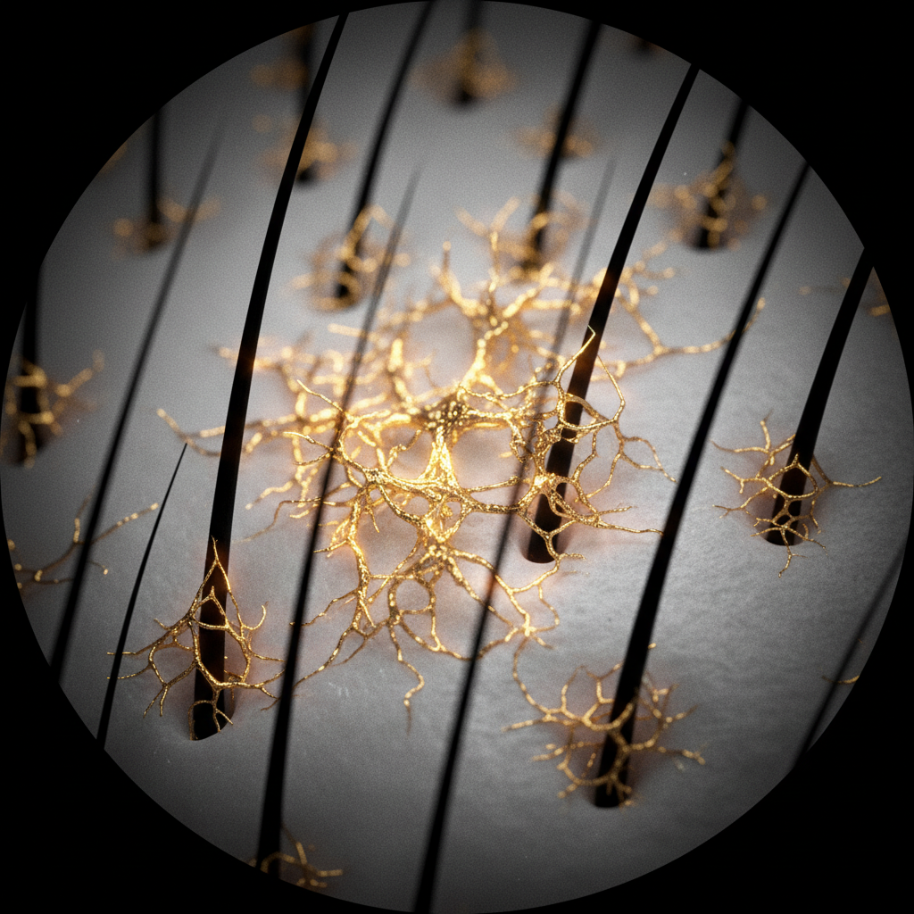
Quick answer: Perifollicular fibrosis explained simply is the microscopic build-up of rigid collagen and scar tissue directly encircling the hair follicle, typically triggered by chronic, low-grade scalp inflammation. As excess connective tissue thickens the follicle sheath, it restricts microcirculation, impedes cellular communication, and mechanically constricts the hair bulb. This progressive stiffening accelerates follicular miniaturisation in pattern hair loss. While advanced scarring cannot be reversed once the follicle stem cell niche is destroyed, early anti-inflammatory scalp care and clinical intervention can halt its progression.
Key takeaways
- Perifollicular fibrosis involves the abnormal deposition of collagen types I and III around the follicular infundibulum and bulge.
- It is most commonly driven by chronic micro-inflammation linked to dihydrotestosterone (DHT), microbial imbalances, or oxidative stress.
- As the fibrous sheath hardens, hair fibres emerge finer and weaker, progressing from terminal hairs to vellus-like strands.
- Fibrosis differs crucially between non-scarring alopecias (where it is mild and perifollicular) and cicatricial alopecias (where it completely obliterates the follicle).
- Topical cosmetics cannot dissolve established scar tissue, but targeted scalp routines can help maintain an optimal environment to discourage inflammatory cascades.
Perifollicular Fibrosis Explained: what the evidence shows
Perifollicular fibrosis refers to excessive extracellular matrix deposition around the hair follicle, predominantly observed in androgenetic alopecia (pattern hair loss) and various cicatricial (scarring) alopecias. Histopathological research shows that this process begins in the upper third of the hair follicle, known as the follicular infundibulum. Rather than being an isolated structural fault, perifollicular fibrosis develops as a downstream consequence of sustained immune system activation and tissue remodelling.
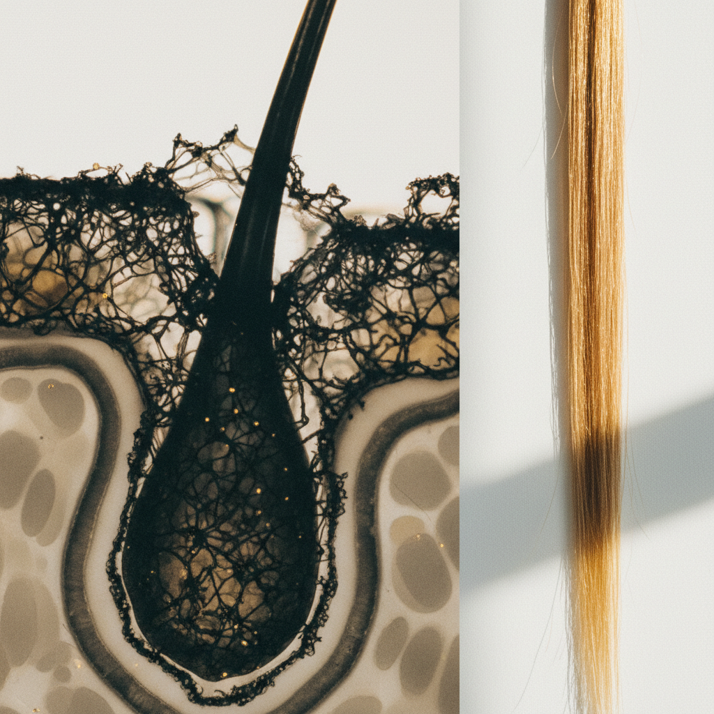
When researchers analyse scalp biopsies from individuals with pattern loss, approximately 30% to 50% demonstrate distinct collagen collar formation surrounding the follicle, alongside an infiltration of activated T-lymphocytes, mast cells, and macrophages. These immune cells release pro-inflammatory cytokines, including transforming growth factor beta (specifically TGF-beta 1), interleukin-1 alpha, and tumour necrosis factor alpha. TGF-beta 1 serves as the master regulator of connective tissue production, prompting dermal fibroblasts to synthesise dense collagen bundles at the expense of healthy, pliable extracellular matrix.
Over time, this fibrous collar creates mechanical resistance against the expanding follicle during the anagen (growth) phase. Instead of anchoring deeply into the subcutaneous fat layer, the follicle becomes trapped in a rigid, fibrotic dermal tract. Consequently, with each hair growth cycle, the hair bulb produces a narrower, shorter shaft, a hallmark of progressive hair thinning.
The biological mechanisms behind follicular stiffening
The biological pathway leading to perifollicular fibrosis involves a tri-part interplay between hormones, inflammatory mediators, and cellular biomechanics:
- Androgen-induced signalling: In genetic pattern hair loss, dihydrotestosterone (DHT) binds to androgen receptors within the dermal papilla cells. This binding does not just shorten anagen; it directly stimulates the secretion of TGF-beta 1 and TGF-beta 2.
- Mast cell activation and cytokine release: Chronic micro-irritation on the scalp surface (often exacerbated by an overgrowth of Malassezia yeasts, lipid peroxides from sebum oxidation, or ultraviolet radiation) triggers mast cells around the follicular infundibulum to degranulate. This releases histamine and pro-inflammatory signalling molecules.
- Fibroblast recruitment and collagen cross-linking: Elevated TGF-beta 1 converts quiescent dermal fibroblasts into active myofibroblasts. These cells produce dense collagenous matrix proteins, primarily type I and type III collagen, while downregulating matrix metalloproteinases (MMPs), the enzymes responsible for breaking down excess scar tissue.
- Vascular compromise and hypoxia: The accumulating connective tissue compresses the microvasculature surrounding the dermal papilla. Reduced blood flow deprives the hair matrix cells of oxygen and micronutrients, accelerating the transition into the catagen (regression) and telogen (resting) phases.
What the evidence does not show
It is critical to dismantle several persistent myths surrounding follicular fibrosis. Laboratory and clinical evidence does not support the idea that everyday physical tension, such as routine brushing or normal styling, causes perifollicular fibrosis. While severe, sustained traction can cause traction alopecia (which may eventually become fibrotic), routine handling does not initiate this cellular cascade.
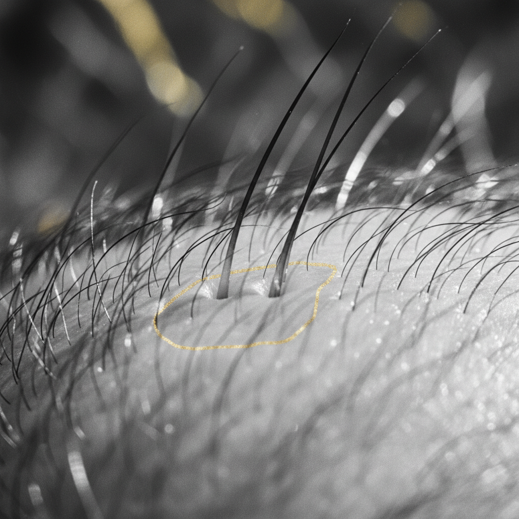
Furthermore, evidence does not show that standard clarifying shampoos, hair oils, or scalp scrubs can "dissolve" or "scrub away" existing collagen bands. Fibrosis occurs deep within the reticular and papillary dermis, well beneath the reach of cosmetic exfoliation. Cosmetic products cannot alter permanent scar tissue, and claims suggesting a shampoo can reverse long-standing structural fibrosis misrepresent the biology of cutaneous wound healing.
Finally, fibrosis is not solely caused by poor circulation. While local hypoxia is an outcome of fibrotic tissue formation, simple vasoconstriction does not spontaneously create collagen deposition without the presence of biochemical inflammatory triggers like TGF-beta.
Key differences: Perifollicular fibrosis across hair conditions
Understanding how fibrosis manifests helps clinicians and stylists differentiate between manageable thinning and irreversible follicle loss.
| Feature | Androgenetic Alopecia (AGA) | Lichen Planopilaris (LPP) | Frontal Fibrosing Alopecia (FFA) |
|---|---|---|---|
| Type of condition | Non-cicatricial (non-scarring) with mild fibrosis | Primary cicatricial (scarring) alopecia | Primary cicatricial (scarring) alopecia |
| Site of fibrosis | Upper sheath, perifollicular connective tissue | Infundibulum and bulge stem cell niche | Bulge stem cell niche and peritrichial rim |
| Inflammatory infiltrates | Low-grade, mast cells and T-lymphocytes | Dense, lichenoid lymphocytic infiltrate | Lymphocytic band-like infiltrate |
| Follicle opening | Present, but often contains smaller vellus hair | Completely lost; smooth, shiny skin | Lost; lone hairs with surrounding pale skin |
| Reversibility | Stabilisation possible; partial recovery of shaft diameter | Irreversible destruction once scarred | Irreversible destruction once scarred |
| Clinical presentation | Gradual thinning on crown, vertex, or temples | Erythema, follicular hyperkeratosis, patchy loss | Receding frontal and temporal hairline, loss of eyebrows |
Signs and symptoms to monitor
Because perifollicular fibrosis develops below the skin's surface, you cannot see the collagen bands directly without a punch biopsy and light microscopy. However, specific scalp and hair signs point to its presence:
- Peripilar signs under dermoscopy: Dermatologists using a trichoscope frequently observe a brown or dark peripilar halo (peripilar sign) measuring roughly 1 millimetre around the follicular opening. This halo correlates histologically with perifollicular inflammation and early fibrotic changes.
- Follicular hyperkeratosis: Tiny, raised collars of keratinised skin around individual hair strands often signal active, low-grade irritation in the upper follicular tract.
- Progressive hair miniaturisation: A visible variation in hair shaft diameters across the scalp (anisotrichosis) indicates that some follicles are being mechanically and biologically constricted by surrounding tissue.
- Scalp dysaesthesia: Persistent, low-level tingling, tenderness, burning, or itchiness around areas of thinning frequently accompanies the release of inflammatory neuropeptides and mast cell degranulation.
What you can do: Supporting the scalp environment
Addressing the factors that contribute to tissue hardening requires an early, multifaceted strategy focusing on calming inflammation, modulating hormonal triggers, and protecting microcirculation.
1. Address surface inflammation promptly
Uncontrolled sebum breakdown and microbial dysbiosis generate free fatty acids that aggravate the follicular infundibulum. Wash your scalp regularly using gentle, pH-balanced formulas (pH 4.5 to 5.5) designed to clear lipid peroxides without stripping the natural barrier.
2. Incorporate botanical and antioxidant support
Certain plant compounds exhibit mild 5-alpha reductase inhibiting and anti-inflammatory properties. Ingredients such as caffeine, rosemary extract, niacinamide, and green tea polyphenols (epigallocatechin gallate) have been documented in laboratory and clinical studies to help mitigate oxidative stress and attenuate TGF-beta signalling pathways in scalp tissue.
3. Consider gentle mechanical stimulation
Evidence suggests that standardised scalp massage, performed for 4 to 5 minutes daily over 24 weeks, alters mechanical strain on dermal papilla cells. In controlled observational trials, this mechanical stimulation increased hair thickness by modulating the expression of genes involved in cellular elasticity and blood flow. However, avoid aggressive or abrasive friction that could induce micro-trauma.
4. Maintain balanced lifestyle factors
Systemic metabolic dysregulation, smoking, and high-glycaemic diets elevate advanced glycation end-products (AGEs). AGEs bind to collagen fibres throughout the body, causing them to become stiff and resistant to standard enzymatic turnover. Eating an antioxidant-rich, balanced diet supports systemic tissue flexibility.
The role of cosmetic haircare and the Watermans approach
Cosmetic haircare cannot replace prescription medical therapies when true scar tissue forms. However, maintaining a clear, calm, and nourished scalp environment is essential for preventing the chronic irritation that feeds early tissue stiffening.
Watermans approaches this balance from a cosmetic and functional perspective. Watermans GrowMe Shampoo is formulated with caffeine, biotin, rosemary extract, and niacinamide to nurture the scalp surface, wash away oxidative sebum build-up, and encourage thicker-, fuller-looking hair. Combined with Watermans ConditionMe Conditioner, which softens the fibre and supports cuticular resilience, this regimen helps reduce breakage while cultivating the clean, calm micro-environment that vulnerable follicles need to thrive.
When the cause may be something else
It is vital to distinguish between true perifollicular fibrosis and other common conditions that do not involve tissue stiffening:
- Telogen effluvium: Characterised by sudden, widespread shedding across the entire scalp 2 to 3 months after a physiological or emotional shock. The follicles remain completely pliable and uninflamed; hair counts rebound once the underlying trigger resolves.
- Hair shaft breakage: Snapping along the hair fibre caused by chemical processing, excessive heat styling, or mechanical friction. The follicle itself remains completely healthy and firmly anchored.
- Seborrhoeic dermatitis: Produces visible yellow, greasy flakes and redness due to yeast sensitivity. While it causes surface inflammation, it rarely generates deep dermal fibrosis unless left completely unmanaged for years.
When to seek professional advice
If you observe rapid hair thinning accompanied by significant scalp burning, pain, scaling, or patches of bare skin that appear smooth and shiny with missing follicle openings, consult a board-certified dermatologist or qualified trichologist immediately.
Primary cicatricial alopecias such as frontal fibrosing alopecia or lichen planopilaris involve aggressive, irreversible fibrosis. These conditions cannot be managed with cosmetic products and require clinical interventions, such as prescription topical or intralesional corticosteroids, calcineurin inhibitors, or systemic immunomodulators to halt permanent destruction of the hair follicle stem cell niche.
Frequently asked questions about Perifollicular Fibrosis Explained
What causes perifollicular fibrosis to develop around hair follicles?
Perifollicular fibrosis is primarily caused by sustained micro-inflammation in the upper follicle. Androgens like DHT trigger the release of transforming growth factor beta 1 (TGF-beta 1), while environmental oxidants, sebum breakdown, and microbial imbalance recruit mast cells and T-lymphocytes. These cells stimulate dermal fibroblasts to produce excessive, rigid collagen bundles that wrap around the follicle.
Can perifollicular fibrosis be completely reversed?
Once dense, mature collagen replaces the hair follicle bulge and destroys the epithelial stem cell reservoir, the damage is permanent. However, if the process is identified in its earliest, non-scarring stages, reducing chronic inflammation and managing DHT levels can halt further fibrosis and help preserve the remaining functional follicles.
How does this guide on Perifollicular Fibrosis Explained help identify scarring alopecia?
This guide on Perifollicular Fibrosis Explained clarifies that while mild fibrosis accompanies regular androgenetic alopecia, scarring (cicatricial) alopecias involve severe, destructive inflammation. If you notice shiny, smooth patches of scalp where follicular pores have disappeared entirely, this indicates cicatricial fibrosis that demands urgent medical evaluation by a dermatologist.
Does scalp massage help prevent tissue hardening?
Gentle, consistent scalp massage can encourage local microcirculation and deliver mechanical stretching forces to dermal papilla cells. Some clinical data suggests this mechanical tension helps stimulate cellular metabolism and decrease the rigidity of local scalp tissue. However, massage should always be gentle to avoid shearing delicate hair fibres or causing additional surface inflammation.
Can cosmetic shampoos cure perifollicular fibrosis?
No cosmetic shampoo can cure or dissolve established dermal scar tissue. Shampoos operate primarily on the surface of the scalp and inside the infundibulum. However, well-formulated products that clear oxidative sebum and deliver soothing ingredients help keep the external environment calm, which discourages the inflammatory triggers associated with tissue stiffening.
Summary
Understanding perifollicular fibrosis explained clearly provides crucial insight into why hair thinning is rarely just a surface cosmetic concern. As chronic micro-inflammation drives the overproduction of stiff collagen collars around hair roots, follicles are progressively compressed, leading to weaker, thinner strands and shortened growth cycles. While advanced, end-stage scarring cannot be undone, intervening early with calming scalp care, lifestyle adjustments, and professional guidance offers the strongest defence in preserving follicle viability and supporting long-term hair density.
Sources and references
- British Association of Dermatologists. Guidelines for the management of alopecia areata and cicatricial alopecias. 2021.
- Jaworsky, C., Kligman, A. M., & Murphy, G. F. Characterization of inflammatory infiltrates in male pattern alopecia. Journal of Investigative Dermatology, 1992.
- Inui, S., & Itami, S. Molecular basis of androgenetic alopecia: From androgen to phenotype. Journal of Dermatological Science, 2013.
- Koyama, T., Kobayashi, K., Hama, T., Murakami, K., & Ogawa, R. Standardized scalp massage results in increased hair thickness by inducing stretching forces to dermal papilla cells in the subcutaneous tissue. Eplasty, 2016.
- Whiting, D. A. Diagnostic and predictive value of horizontal sections of scalp biopsy specimens in male pattern androgenetic alopecia. Journal of the American Academy of Dermatology, 1993.
Find these ingredients in Watermans products
- Niacinamide: you will find this ingredient in our Hair Mask.