Understanding Frontal Fibrosing Alopecia: Causes, Signs, and Management
8 min read ·
Understanding Frontal Fibrosing Alopecia is vital for spotting early signs of this scarring condition, preserving remaining hair, and getting timely medical guidance.

Quick answer: Understanding Frontal Fibrosing Alopecia is essential for identifying this progressive form of primary cicatricial (scarring) hair loss, which primarily affects postmenopausal women along the frontal and temporal hairline. It causes irreversible destruction of the hair follicle stem cells via immune-mediated inflammation, often leading to eyebrow loss and a smooth band of receded skin. The main limitation is that lost follicles cannot be regenerated, meaning clinical management focuses strictly on halting progression rather than regrowing hair.
Key takeaways
- Frontal Fibrosing Alopecia (FFA) is a lymphocytic scarring alopecia considered a clinical variant of lichen planopilaris.
- The condition causes permanent follicle destruction, replacing active follicular openings with fibrous scar tissue.
- Early warning signs include symmetrical hairline recession, loss of eyebrows, redness (erythema), and scaling (perifollicular hyperkeratosis) around follicles.
- Topical cosmetic products and regular hair care cannot cure or halt FFA, which requires prescription anti-inflammatory dermatological management.
- Gentle, low-tension styling and neutral scalp hygiene help protect vulnerable neighbouring fibres from physical breakage and secondary irritation.
Understanding Frontal Fibrosing Alopecia: what the evidence shows
Understanding Frontal Fibrosing Alopecia requires examining its classification as a primary cicatricial alopecia. First described in medical literature by Australian dermatologist Steven Kossard in 1994, the condition has seen an unexplained surge in diagnoses globally since the early 2000s.
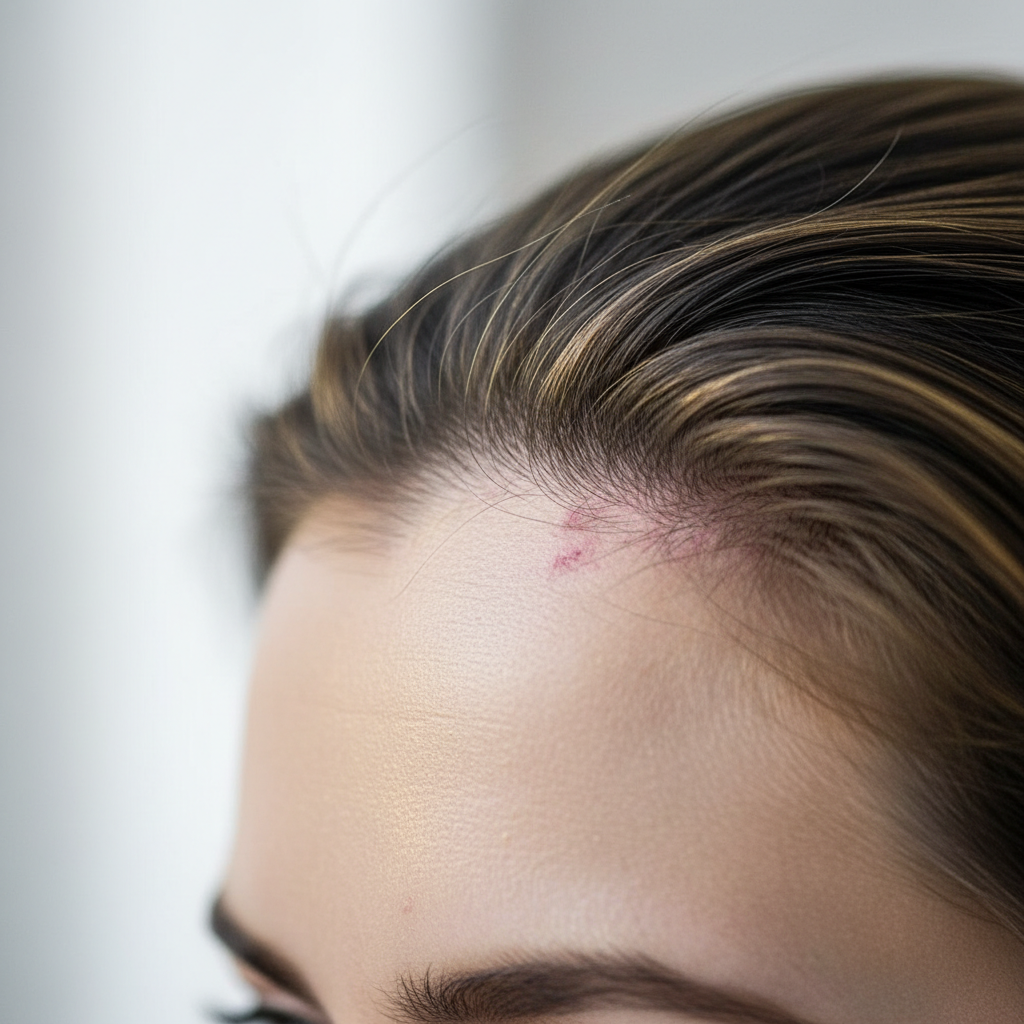
Clinical research establishes that FFA is driven by an autoimmune lymphocytic attack targeting the upper region of the hair follicle, specifically the infundibulum and isthmus. This area houses critical epithelial stem cells responsible for cycling and regenerating the follicle. When cytotoxic T-lymphocytes infiltrate this zone, they trigger chronic inflammation that destroys the sebaceous glands and stem cell niche. As healing takes place, the body replaces the complex follicular apparatus with fibrous connective tissue, permanently extinguishing the hair follicle's ability to produce a fibre.
Epidemiological studies indicate that roughly 85 to 90 percent of diagnosed individuals are postmenopausal women, typically presenting between the ages of 55 and 65. However, dermatologists increasingly diagnose premenopausal women, and approximately 2 to 5 percent of reported cases occur in men. The exact trigger remains actively investigated, with current evidence pointing toward an interaction between genetic susceptibility (such as human leukocyte antigen alleles), hormonal shifts, and potential environmental or chemical exposures.
What the evidence does not show: myths and misconceptions
Despite increased research over the past two decades, several misconceptions persist regarding the triggers and behaviour of the condition:
- Sunscreen as a direct cause: Several observational studies noted an association between daily facial sunscreen use and FFA. However, systemic reviews by dermatological associations clarify that correlation does not prove causation. Sunscreen use may simply reflect heightened facial care regimens or skin sensitivity in this demographic. Major dermatology bodies do not recommend discontinuing sun protection.
- Cosmetics as the primary culprit: Ordinary hair dyes, shampoos, and conditioners do not cause the internal immune assault that drives FFA.
- Guaranteed total baldness: FFA typically stabilises after progressing across a variable band, usually between 1 to 5 centimetres of recession, though complete vertex involvement is rare.
- Reversibility with diet: Dietary interventions, vitamins, or nutritional supplements have no established clinical track record of reversing the follicular scarring of FFA.
Signs and clinical presentation of FFA
Recognising FFA early is critical because medical intervention cannot revive scarred tissue; it can only protect existing follicles. The presentation typically features several distinct clinical markers:
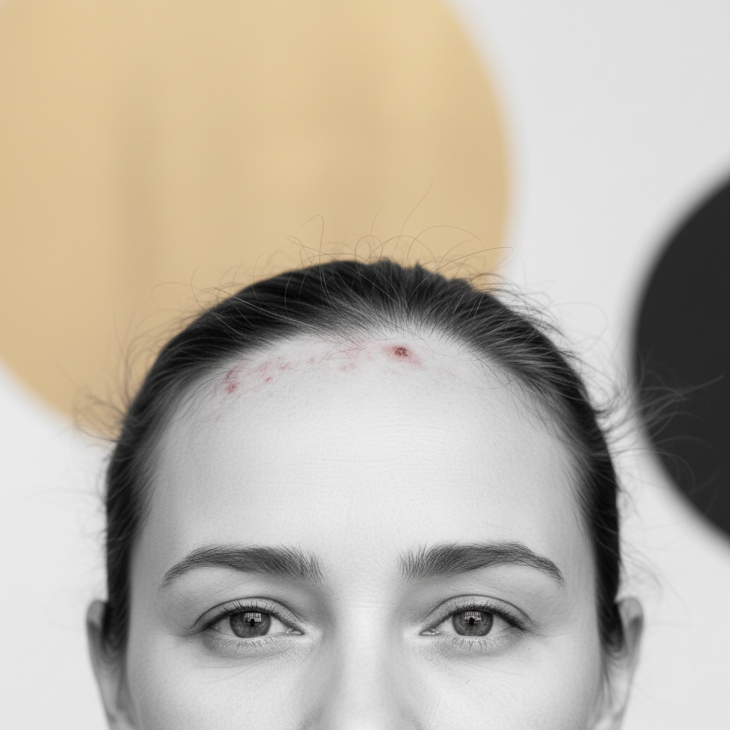
- Marginal hairline recession: A uniform, symmetrical band of hair loss appears along the frontal hairline and temporal peaks, leaving the exposed forehead skin remarkably smooth, pale, and devoid of visible follicular pores.
- The "lonely hair" sign: Isolated, single terminal hairs frequently persist within the completely bald, receded frontal band.
- Eyebrow thinning (madarosis): Eyebrow loss occurs in up to 80 percent of patients. In many individuals, the outer third of the eyebrows thins or disappears entirely months or years before hairline recession becomes obvious.
- Perifollicular erythema and hyperkeratosis: Active margins of the hairline often display tiny rings of redness (erythema) and small collarettes of dry, rough scale adhering to the base of individual hairs.
- Facial papules: Non-inflammatory, skin-coloured or yellowish bumps can appear on the temples and cheeks, representing involvement of facial vellus follicles.
- Scalp sensations: Patients frequently report burning, tenderness, tingling (trichodynia), or severe itching along the front margin of the scalp.
Distinguishing FFA from other conditions
Because the frontal hairline is prone to multiple types of hair loss, misdiagnosis is common during early stages.
| Feature | Frontal Fibrosing Alopecia (FFA) | Traction Alopecia | Female Pattern Hair Loss (FPHL) |
|---|---|---|---|
| Primary Category | Scarring (cicatricial) alopecia | Mechanical hair loss | Non-scarring progressive miniaturisation |
| Follicular Openings | Absent; skin looks shiny and smooth | Retained initially; lost only in late stages | Fully preserved throughout |
| Eyebrow Involvement | Frequent (up to 80% of cases) | Rare or absent | Absent |
| Scalp Appearance | Perifollicular erythema, scaling collarettes | Fringe sign, broken hairs, folliculitis | Normal scalp skin, no scarring |
| Sensation | Common itching, burning, pain | Mild tenderness following tight styling | Typically asymptomatic |
| Reversibility | Irreversible once scarred | Fully reversible if caught early | Miniaturised hairs can be enlarged |
What you can do: clinical management and cosmetic care
Managing Frontal Fibrosing Alopecia involves two distinct branches: prescription medical therapy administered by a dermatologist, and supportive cosmetic maintenance to prevent mechanical damage to existing strands.
Dermatological medical interventions
Dermatologists aim to extinguish the active T-cell inflammation as quickly as possible. Standard clinical approaches documented in clinical guidelines include:
- Intralesional corticosteroids: Targeted injections (such as triamcinolone acetonide) along the active inflammatory margin to rapidly damp down localized inflammation.
- Topical anti-inflammatories: High-potency topical corticosteroids and topical calcineurin inhibitors (tacrolimus or pimecrolimus) to calm perifollicular redness and burning.
- Systemic immunomodulators: Oral medications such as hydroxychloroquine, tetracycline-class antibiotics (for their anti-inflammatory properties), or 5-alpha reductase inhibitors (finasteride or dutasteride), which have shown utility in slowing progression in clinical cohorts.
Supportive daily hair and scalp practices
While cosmetics cannot arrest scarring, harsh handling accelerates breakage on already compromised hair:
- Zero-tension styling: Avoid pulling the hair backward into tight ponytails, buns, or headbands. Any traction adds mechanical stress to inflamed follicles.
- Mild, pH-balanced cleansing: Wash the scalp regularly with a gentle shampoo (formulated around pH 5.0 to 5.5) to clear secondary sebum build-up and environmental grime without irritating the barrier.
- Gentle detangling: Comb hair from tips to roots using wide-tooth implements, never yanking against snags.
- Minimise heat: Keep blow dryers on cool or warm settings, directing airflow away from the bare frontal skin.
When to seek specialist evaluation
If you notice your frontal hairline creeping backward, an uncharacteristic loss of your outer eyebrows, or persistent redness and itchiness at the hair margins, request an immediate referral to a consultant dermatologist.
A dermatologist will perform trichoscopy (dermoscopy of the scalp and hair). Under magnification, the characteristic signs of FFA, including absent follicular ostia, peripilar casts, and subtle perifollicular erythema, are easily visible to a trained eye. In ambiguous cases, a 4 mm punch biopsy taken from the active inflammatory border can confirm the diagnosis histopathologically, allowing targeted treatment before additional follicles undergo irreversible fibrotic replacement.
Supporting hair condition: Watermans scalp philosophy
When managing conditions that affect scalp comfort, maintaining a clean, balanced environment supports overall cosmetic hair density. Watermans focuses on nurturing the appearance of natural hair through revitalising topical formulas that fortify the hair shaft against breakage.
While Watermans products do not treat, cure, or arrest Frontal Fibrosing Alopecia or any other form of medical cicatricial scarring, maintaining an optimal scalp foundation is beneficial for non-scarred hair. Formulas such as Watermans Grow Me Shampoo incorporate nourishing ingredients like biotin, rosemary, caffeine, and niacinamide. These cosmetic agents help wash away scalp residues, support skin moisture, and condition the surviving hair lengths so they look fuller, thicker, and more resilient against daily wear.
Frequently asked questions about Understanding Frontal Fibrosing Alopecia
What is the primary difference between alopecia areata and Frontal Fibrosing Alopecia?
Alopecia areata is an autoimmune, non-scarring condition that typically causes sudden, round, smooth patches across the scalp or body while preserving the underlying follicular structures, meaning hair can fully regrow. In contrast, Frontal Fibrosing Alopecia is a cicatricial (scarring) condition that permanently destroys the follicle and stem cells, resulting in irreversible hairline recession.
Can hair grow back once Frontal Fibrosing Alopecia has caused a bare patch?
No. Once a follicle has undergone the fibrotic replacement characteristic of Frontal Fibrosing Alopecia, the follicle is replaced by scar tissue and cannot generate a new fibre. This makes early clinical recognition and prompt medical treatment vital to halt the process and preserve the remaining hair.
Is hair transplantation an effective option for FFA?
Hair transplantation into areas cleared by FFA carries significant risk. While transplants can initially survive, published surgical studies reveal that the underlying lichenoid inflammation often recurs, destroying the newly transplanted grafts within 2 to 5 years. Dermatologists usually recommend waiting until the condition has been completely quiescent and inactive for at least two years before considering surgical options.
Does colouring or dying hair trigger or worsen Frontal Fibrosing Alopecia?
There is no reproducible medical evidence showing that chemical hair dyes trigger or exacerbate the autoimmune mechanism of FFA. However, applying harsh chemical treatments directly onto an actively inflamed, tender scalp can cause acute contact dermatitis or chemical irritation, increasing discomfort and contributing to shaft breakage.
Effective management and outlook
Understanding Frontal Fibrosing Alopecia empowers you to seek early, proactive medical intervention before extensive hairline loss occurs. Because FFA is a progressive, scarring condition that permanently destroys follicles, cosmetic remedies alone are insufficient; working alongside a dermatologist offers the best pathway to halt inflammation and preserve your natural hairline. By pairing targeted medical care with gentle, tension-free hair maintenance, you can protect your remaining hair and support long-term scalp health.
Sources and references
- Kossard, S. (1994). Postmenopausal frontal fibrosing alopecia: Scarring alopecia in a pattern distribution. Archives of Dermatology, 130(6), 770-774.
- British Association of Dermatologists. (2020). Frontal Fibrosing Alopecia: Clinical Guidelines and Management.
- Vañó-Galván, S., et al. (2014). Frontal fibrosing alopecia: a multicenter review of 355 patients. Journal of the American Academy of Dermatology, 70(4), 670-678.
- Harries, M. J., et al. (2018). Lichen planopilaris and frontal fibrosing alopecia: an overview of pathogenesis and clinical presentation. British Journal of Dermatology, 179(4), 819-826.
- National Institute for Health and Care Excellence (NICE). Alopecia - cicatricial: Clinical knowledge summaries.
Find these ingredients in Watermans products
- Niacinamide: you will find this ingredient in our Hair Mask.