Trichoscopy Essentials for Scalp Health: The Clinical Guide
9 min read ·
Master Trichoscopy Essentials for Scalp Health to identify hair diameter diversity, follicular dots, and early hair loss signs with microscopic clinical clarity.
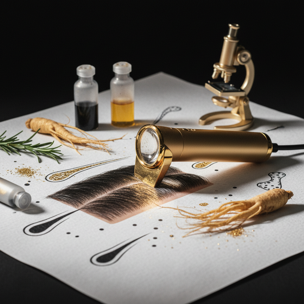
Quick answer: Understanding Trichoscopy Essentials for Scalp Health allows clinicians, trichologists, and stylists to examine the hair and scalp non-invasively at 10x to 70x magnification. By evaluating follicular openings, hair shaft diameter diversity, perifollicular skin, and vascular patterns, trichoscopy distinguishes between non-scarring conditions like androgenetic alopecia and scarring cicatricial alopecias early. Its main limitation is that while it reveals distinct structural patterns, it cannot replace a histopathological scalp biopsy when inflammatory scarring conditions present with ambiguous or overlapping features.
Key takeaways
- Trichoscopy is dermoscopy of the hair and scalp, offering rapid, non-invasive magnification ranging from 10x up to 70x using polarised or non-polarised light.
- Diagnostic clues fall into four core visual categories: follicular openings (dots), hair shaft morphology, perifollicular epidermis, and microvascular structures.
- Hair diameter diversity exceeding 20 percent across terminal hairs is the classic dermoscopic hallmark of androgenetic alopecia.
- Yellow dots suggest sebum retention in distended follicles, while black dots indicate cadaverised hairs snapped at the scalp surface.
- Early identification of scarring alopecias (such as lichen planopilaris) protects against irreversible follicular destruction and permanent hair loss.
- Cosmetic scalp care can improve surface hydration and microcirculation, but clinical trichoscopy findings require qualified dermatological assessment.
Trichoscopy Essentials for Scalp Health: what the evidence shows
Trichoscopy relies on reflected-light microscopy to make sub-surface structures visible through the epidermis without surgical excision. Peer-reviewed dermatological studies confirm that trichoscopy significantly increases diagnostic accuracy for hair and scalp disorders compared to unaided visual inspection.

When dermatologists evaluate a patient using trichoscopy, they examine four primary parameters across standard reference zones: the frontal hairline, the vertex, the parietal ridges, and the occipital scalp.
1. Follicular openings and dots
Follicular dots represent the openings of hair follicles (ostia) viewed from above:
- Yellow dots: Follicular infundibula filled with sebaceous material and keratin debris. They appear prominently in alopecia areata, but can also be seen in severe androgenetic alopecia.
- Black dots (cadaverised hairs): Pigmented hairs that have fractured flush with the scalp surface before emergence. They are characteristic of acute, highly active alopecia areata, trichotillomania, and certain fungal infections (tinea capitis).
- White dots: Can indicate pinpoint physiological sweat gland duct openings, or fibrotic tissue (follicular scarring) where the follicular unit has been permanently replaced by collagen, typical in cicatricial alopecias.
- Red dots: Erythematous follicular centres often tied to discoid lupus erythematosus.
2. Hair shaft characteristics
Healthy scalp hair displays relatively uniform thickness across terminal fibres, with an average diameter between 0.06 mm and 0.08 mm. Trichoscopy reveals several shaft abnormalities:
- Anisotrichosis (hair diameter diversity): Variation in hair shaft thickness greater than 20 percent across a single field of view is the gold-standard trichoscopic indicator of follicle miniaturisation in androgenetic alopecia.
- Exclamation mark hairs: Short broken hairs (1 to 3 mm long) that are thinner at the proximal base near the scalp and thicker distally. These indicate active follicular inflammation in alopecia areata.
- Broken hairs and trichorrhexis nodosa: Fractured fibres displaying frayed, brush-like tips, pointing toward chemical, mechanical, or thermal damage rather than root-level pathology.
- Pigtail hairs and circle hairs: Short, circularly regrowing hairs often observed during remission in shedding disorders.
3. Perifollicular epidermis and scaling
The skin immediately surrounding the follicular exit provides crucial information about inflammation and epidermal turnover:
- Peripilar casts (perifollicular collars): Tubular white-silver scales that wrap around the proximal hair shaft. Dense tubular casts that do not move freely along the shaft signal lichen planopilaris or frontal fibrosing alopecia.
- Interfollicular scaling: Diffuse, greasy, yellowish scales suggest seborrhoeic dermatitis, whereas thick, silvery, adherent plaques point to scalp psoriasis.
- Honeycomb pigment pattern: A physiological, reticular brown pigment network seen on sun-exposed, thinned scalps, particularly in individuals with higher phototypes.
4. Microvascular patterns
Normal scalp microvasculature presents as simple red loops or arborising (branching) red lines. Twisted red loops are characteristic of scalp psoriasis, while thin, tortuous, branching vessels surrounded by pale halos can reflect active chronic discoid lupus or dermatomyositis.
| Trichoscopic Feature | Common Finding / Morphology | Associated Condition | Clinical Meaning |
|---|---|---|---|
| Hair Diameter Diversity | >20% variation in shaft thickness | Androgenetic Alopecia | Progressive follicular miniaturisation |
| Yellow Dots | Round, amorphous yellowish structures | Alopecia Areata, Androgenetic Alopecia | Sebaceous and keratin pooling in empty/miniaturised ostia |
| Exclamation Mark Hairs | Short hairs, tapered at proximal base | Alopecia Areata | Acute autoimmune insult to hair matrix |
| Peripilar Tubular Casts | Dense white keratin sleeves wrapping hair shaft | Lichen Planopilaris, Frontal Fibrosing Alopecia | Follicular infundibular inflammation (scarring risk) |
| Black Dots | Pigmented remnants flush with ostium | Alopecia Areata, Tinea Capitis | Rapid hair fracture within the infundibulum |
| Twisted Red Loops | Concentrated microvascular hairpin turns | Scalp Psoriasis | Accelerated epidermal proliferation and angiogenesis |
What the evidence does not show
Trichoscopy cannot replace histopathology in every clinical scenario. While a dermatoscope exposes micro-features invisible to the naked eye, it cannot definitively verify cellular infiltrate profiles or reveal deep subcutaneous pathology without a 4 mm punch biopsy.
Furthermore, trichoscopy cannot diagnose the systemic root causes of telogen effluvium. An acute diffuse telogen shed will often show empty follicles and short upright regrowing hairs under magnification, but the dermatoscope cannot determine whether the underlying cause is an iron deficiency, thyroid dysfunction, severe psychological stress, or postpartum hormonal shifts. Blood biochemistry panels remain mandatory.
Trichoscopy tools marketed for home use also have limitations. Without extensive training in dermatological diagnostic criteria, high-magnification smartphone attachments frequently lead to misinterpretation, mistaking ordinary sebum deposits for serious disease or normal variation for pathology.
Mechanisms: how magnification reveals follicular dynamics
The human scalp contains approximately 100,000 hair follicles, clustered naturally in follicular units of one to four terminal hairs, accompanied by one or two vellus hairs, sebaceous glands, and a shared erector pili muscle.
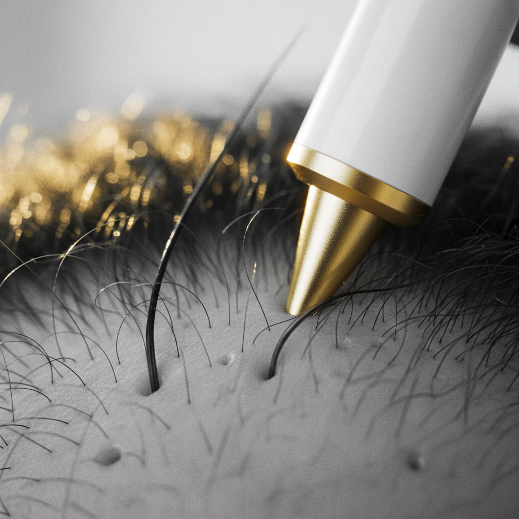
Under normal physiology, the ratio of terminal to vellus hairs on the scalp is roughly 7 to 1 or higher. In androgenetic alopecia, dihydrotestosterone (DHT) binds to androgen receptors in genetically susceptible dermal papilla cells. This binding shortens the anagen (growth) phase and prolongs the telogen (resting) phase, progressively shrinking the hair follicle.
Using polarised trichoscopy, clinicians assess this phenomenon directly. Polarisation eliminates surface skin glare without requiring interface fluid (such as ultrasound gel or alcohol), allowing light to penetrate into the upper dermis.
By measuring the exact proportion of miniaturised vellus-like hairs (diameter under 0.03 mm) compared to robust terminal hairs, practitioners quantify follicular decline long before visible thinning or widened partings appear on global clinical photography.
Signs to look for: clinical vs cosmetic observations
Salon professionals and trichologists often observe early changes during washing, cutting, and scalp consultations:
- Surface tightness and redness: Visible pink or violaceous hues around individual follicles warrant attention, as early folliculitis or scarring conditions begin with subtle perifollicular erythema.
- Clustered missing follicles: If skin appears unusually shiny, pale, and entirely devoid of follicular openings, this indicates follicular dropout, which requires immediate medical referral.
- Excessive shed bulbs: Differentiating between white keratin bulbs (telogen shedding) and dystrophic, sheared ends (mechanical breakage) helps determine whether the issue stems from physiological cycles or structural fibre damage.
What you can do to support the scalp environment
While medical conditions require targeted medical treatment, creating an optimal external environment helps support normal follicular cycling:
- Maintain a balanced scalp pH: Healthy scalp skin maintains an acidic pH between 4.5 and 5.5, which preserves the acid mantle and deters pathogenic microbes like excessive Malassezia yeast.
- Cleanse systematically: Infrequent washing allows sebum, squalene peroxides, and dead corneocytes to accumulate within follicular funnels, creating yellow dot congestion and triggering inflammatory cascades.
- Avoid high-friction styling: Excessive tension from tight ponytails, weaves, or aggressive brushwork can induce mechanical stress, culminating in traction alopecia, visible on trichoscopy as peripilar casts and hair cast formation along the marginal hairline.
The role of supportive cosmetic care
When underlying pathology has been ruled out or is being managed by a clinician, high-quality cosmetic formulations can support the appearance of hair density and scalp comfort.
Watermans Grow Me Shampoo and Condition Me Conditioner provide a gentle, sulfate-free approach that thoroughly cleanses sebum and debris from follicular openings without disrupting the epidermal moisture barrier. Formulated with targeted cosmetic ingredients including biotin, caffeine, rosemary extract, niacinamide, and argan oil, the range is designed to revitalise the scalp surface, nourish the hair fibre, and reduce breakage so existing hair looks fuller and healthier.
When to seek professional medical advice
Immediate referral to a dermatologist or qualified medical trichologist is required if trichoscopy or visual inspection reveals:
- Rapidly expanding, patchy hair loss with smooth bare skin.
- Symptoms of burning, tenderness, pruritus, or pain localized to the scalp.
- Pustules, crusting, or bleeding around follicular units.
- Progressive recession of the frontal or temporal hairline accompanied by eyebrow thinning.
- Complete loss of follicular ostia (smooth, porcelain-like ivory patches).
Frequently asked questions about Trichoscopy Essentials for Scalp Health
How does trichoscopy differ from standard dermoscopy?
Trichoscopy is dermoscopy applied specifically to the scalp and hair. While standard dermoscopy focuses primarily on evaluating pigmented skin lesions and melanomas across the body, trichoscopy concentrates on follicular openings, perifollicular skin, hair shaft morphology, and scalp vascular patterns to diagnose hair disorders.
Can Trichoscopy Essentials for Scalp Health replace a scalp biopsy?
Trichoscopy essentials for scalp health significantly reduce the need for scalp biopsies by providing non-invasive diagnostic clues. However, trichoscopy cannot completely replace a histopathological biopsy when scarring alopecias present with atypical features, mixed signs, or ambiguous microscopic margins.
How often should a trichoscopy assessment be repeated?
For monitoring treatment progress in conditions like androgenetic alopecia or alopecia areata, trichoscopy is typically repeated every 3 to 6 months. This timeframe allows sufficient anagen hair growth to accurately measure changes in hair density, shaft calibre, and follicular regrowth.
Can a trichoscopy exam identify hair breakage versus hair shedding?
Yes. Trichoscopy clearly distinguishes hair breakage from shedding. Shedding reveals empty follicular openings or hairs exiting normally with preserved tips, whereas breakage displays frayed ends, trichorrhexis nodosa nodes, or abrupt fracture margins along the hair shaft without empty follicular ostia.
Trichoscopy Essentials for Scalp Health in modern practice
Mastering Trichoscopy Essentials for Scalp Health bridges the gap between basic visual consultation and microscopic precision. By methodically categorising hair shaft diversity, follicular dots, perifollicular scale, and microvasculature, practitioners can identify early signs of miniaturisation and active inflammation.
Early detection remains the single most effective tool for preventing irreversible follicle loss. Incorporating routine dermoscopic evaluation into scalp care ensures that subtle signs are addressed promptly, protecting long-term hair density and scalp health.
Sources and references
- British Association of Dermatologists. Guidelines for the management of alopecia areata. Published 2012, updated 2020.
- Rudnicka, L., Olszewska, M., Rakowska, A., & Slowinska, M. Atlas of Trichoscopy: Dermoscopy in Hair and Scalp Disease. Springer, 2012.
- Miteva, M., & Tosti, A. Hair and scalp dermatoscopy. Journal of the American Academy of Dermatology, 2012. DOI: 10.1016/j.jaad.2012.02.013.
- National Institute for Health and Care Excellence (NICE). Alopecia androgenetica: Clinical Knowledge Summaries. Updated 2022.
- Rossi, A., et al. Trichoscopy in the diagnosis of hair and scalp disorders. Dermatologic Clinics, 2018.
Find these ingredients in Watermans products
- Niacinamide: you will find this ingredient in our Hair Mask.