Trichoscopy Basics: The Essential Guide to Scalp Dermoscopy
9 min read ·
Learn trichoscopy basics to accurately assess hair follicles, scalp patterns, shaft breakage, and shedding using non-invasive dermoscopic magnification.
Quick answer: Understanding Trichoscopy Basics allows clinicians and hair professionals to examine the scalp and hair fibres using high-magnification dermoscopy (typically 10x to 70x or higher). It visualises follicular openings, perifollicular skin, hair shaft calibre, and vascular patterns without taking a tissue biopsy. The primary limitation of trichoscopy is that while it accurately identifies structural patterns and inflammatory signs, it cannot replace histological biopsy or blood pathology when diagnosing atypical, overlapping, or early-stage scarring alopecias.
Key takeaways
- Trichoscopy is non-invasive, bedside dermoscopy specifically adapted for hair and scalp disorders.
- It evaluates four core anatomical features: follicular openings (dots), perifollicular epidermis, hair shaft morphology, and vascular structures.
- The technique separates non-scarring alopecias (where follicular openings remain intact) from scarring alopecias (where follicular openings are permanently lost).
- Handheld dermoscopes provide 10x magnification, whereas digital videodermoscopes can magnify structures up to 70x to 100x.
- Trichoscopy cannot evaluate systemic biochemical causes, such as ferritin deficiencies or thyroid imbalances, which still require laboratory testing.
Trichoscopy Basics: what the evidence shows
Trichoscopy represents the intersection of dermatology, optical magnification, and trichology. Peer-reviewed dermatological literature confirms that examining the scalp under polarised and non-polarised light magnifies features invisible to the naked human eye.
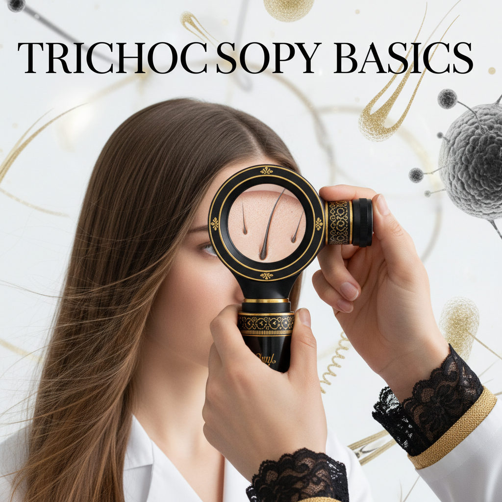
During an examination, a clinician places a dermatoscope directly against the scalp, using either dry contact, cross-polarised illumination, or an interface liquid (such as 70% alcohol or ultrasound gel) to reduce surface light reflection. Polarised light penetrates deeper into the dermis to illuminate vascular structures and collagen, while non-polarised light with immersion fluid gives crisp resolution of the superficial epidermal surface and follicular ostia.
Clinical studies establish that healthy scalp skin displays uniform hair shaft thickness (with less than 20% diameter diversity) and follicular units predominantly containing two to four terminal hairs. When pathology develops, this architecture shifts in measurable ways. Trichoscopy allows practitioners to catch these shifts months before gross thinning becomes visible to the naked eye.
The four core structures evaluated in trichoscopy
To master the foundations of scalp analysis, clinicians systematically review four distinct visual components.
1. Follicular openings (dots)
The openings where hair shafts emerge are known clinically as ostia. Their presence, colour, and contents provide immediate diagnostic clues:
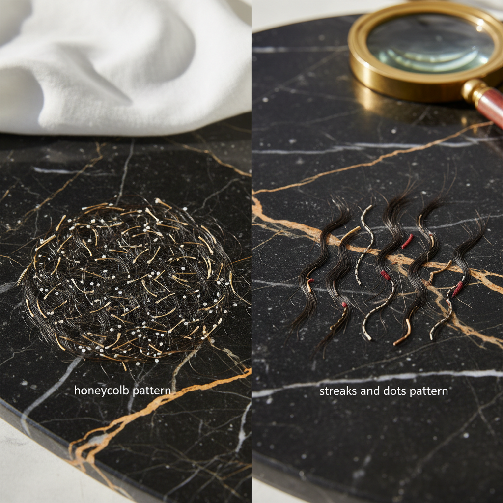
- Yellow dots: Follicular infundibula distended with sebum and keratinous debris. They are classical hallmarks of alopecia areata, but can also appear in severe androgenetic alopecia (AGA).
- Black dots: Cadaverised hairs. These form when pigmented hair fibres fracture at the level of the scalp surface due to rapid, acute follicular damage, commonly seen in active alopecia areata or tinea capitis.
- White dots: These represent either pinpoint fibrous tracts replacing destroyed follicles in primary scarring alopecias (such as lichen planopilaris), or empty sweat gland ducts against deeply pigmented skin.
- Red dots: Erythematous circles surrounding follicles, frequently linked to active discoid lupus erythematosus.
2. Hair shaft signs
Trichoscopy allows detailed calibre analysis along the hair shaft:
- Hair diameter diversity (anisotrichosis): A variation in hair shaft thickness greater than 20% is the primary diagnostic criterion for androgenetic alopecia. It reflects gradual follicular miniaturisation.
- Exclamation mark hairs: Short broken hairs (typically 1 to 4 mm long) that are distinctly thinner at the proximal base than at the distal tip. These indicate an abrupt interruption of cortical keratinisation, classic in active alopecia areata.
- Pigtail or circle hairs: Tightly coiled short regrowing hairs that signal early recovery or rapid cycling.
- Monilethrix and trichorrhexis nodosa: Specific structural genetic or acquired shaft breakages, seen as regular beaded nodes or brush-like fractures under magnification.
3. Perifollicular skin
The epidermis immediately surrounding the follicle base reveals the presence and nature of inflammation:
- Peripilar casts (perifollicular scales): Tubular collars of keratin wrapped around the base of the hair shaft. White tubular scaling strongly indicates lichen planopilaris or frontal fibrosing alopecia, whereas greasy, yellowish flakes clinging loosely to the scalp point to seborrhoeic dermatitis.
- Peripilar sign: A brown or dark halo (around 1 mm in diameter) encircling the follicular opening. This indicates superficial perifollicular lymphocytic inflammation, common in early-stage androgenetic alopecia.
4. Vascular patterns
Under polarised light, the microvasculature of the superficial dermis becomes apparent:
- Simple red loops and arborising red lines: Normal microcirculation often looks like faint, uniform hairpin loops.
- Arborising vessels (branching tree-like vessels): Prominent in discoid lupus erythematosus and basal cell carcinoma of the scalp.
- Interfollicular red networks: Distinct patterns common in scalp psoriasis, often showing bushy, twisted capillary loops, contrasting with the fine regular loops of seborrhoeic dermatitis.
Diagnostic patterns compared
| Condition | Follicular Openings | Hair Shaft Findings | Scalp Surface / Vascular Signs |
|---|---|---|---|
| Androgenetic Alopecia (AGA) | Yellow dots (in advanced stages); empty ostia | Anisotrichosis (>20% diversity); miniaturised vellus hairs | Peripilar brown halo; single-hair follicular units dominate |
| Telogen Effluvium (TE) | Normal openings; occasionally empty ostia | Uniform thickness (anisotrichosis <20%); short upright regrowing hairs | No perifollicular inflammation; normal vascular loops |
| Alopecia Areata (AA) | Yellow dots; black dots (cadaverised hairs) | Exclamation mark hairs; micro-exclamation hairs; tapered hairs | Normal interfollicular skin; no loss of follicular ostia |
| Lichen Planopilaris (LPP) | Complete loss of ostia in scarred patches; white dots | Absence of vellus hairs; occasional cast hairs | Perifollicular erythema; hyperkeratotic white peripilar casts |
| Scalp Psoriasis | Normal ostia beneath scales | Normal shaft morphology; no focal miniaturisation | Thick silvery scales; bushy, tortuous capillary loops |
What the evidence does not show
While trichoscopy is a powerful clinical aid, its capabilities have defined scientific boundaries.
Trichoscopy cannot confirm the biochemical, nutritional, or endocrine drivers of hair shedding. It cannot measure serum ferritin, vitamin D3, thyroid-stimulating hormone (TSH), or free androgen levels. A client showing uniform telogen shedding will display relatively normal trichoscopic features; the scope cannot differentiate between post-febrile shedding, postpartum shedding, or nutritional restriction.
Furthermore, trichoscopy cannot always provide a definitive verdict in ambiguous, early cicatricial (scarring) conditions. Early frontal fibrosing alopecia or traction alopecia can present with overlapping features. When follicular destruction is suspected, dermoscopy pinpoints the ideal, active edge of a lesion to perform a punch biopsy, but it does not replace the subsequent histological examination.
Differentiating hair shedding from hair breakage
One of the most valuable day-to-day applications of trichoscopy is resolving the confusion between shedding and mechanical breakage. Clients frequently arrive at salons or clinics reporting that their hair is falling out from the roots, when in reality the hair shaft is snapping mid-length.
- Shedding (telogen release): Trichoscopy reveals clean, open follicular ostia, an absence of fractured stumps, and normal hair fibre architecture. A gentle pull test evaluated under magnification shows hair fibres with intact, club-shaped keratinised bulbs at the proximal end.
- Breakage (mechanical or chemical trauma): The scalp reveals normal follicle density, but high-magnification trichoscopy detects fraying, transverse fractures (trichorrhexis nodosa), split ends (trichoptilosis), and irregular shaft lengths without club roots. This is common following aggressive chemical processing, excessive heat styling, or heavy tension styling.
What you can do: clinical and salon workflow
For hair professionals, stylists, and clinic practitioners incorporating trichoscopic imaging into consultations, follow this structured routine:
- Systematic mapping: Check at least four zones: frontal hairline, vertex, temporal regions, and occiput. The occiput serves as an androgen-independent control area to assess your client's baseline hair thickness and density.
- Clean scalp assessment: Ideally, perform the examination on hair that has not been washed for 24 to 48 hours to assess sebum and scaling, but is free from dry shampoos, fibres, or heavy styling waxes that mimic black or yellow dots.
- Calibre check: Measure hairs within a single field of view. If more than one in five hairs is noticeably thinner than surrounding hairs, document this as anisotrichosis.
- Photodocumentation: Store calibrated macro photographs of standard focal spots. Repeat imaging at 3 to 6 month intervals provides objective evidence of hair preservation or progression.
Supporting the scalp environment
Cosmetic topicals cannot reverse genetic follicle miniaturisation or repair fibrosed, scarred tissue. However, evidence shows that maintaining a clear, non-inflamed scalp environment optimizes the conditions needed for healthy growth cycles.
Formulations like Watermans GrowMe shampoo and condition-focused topicals use cosmetic ingredients such as biotin, rosemary, caffeine, and niacinamide. These cosmetic preparations help keep follicular ostia clear of oxidised sebum, reduce surface flaking, and condition the hair fibre to minimise breakage along the shaft. When paired with routine trichoscopic monitoring, this supportive approach ensures external factors (like heavy cosmetic buildup and mechanical snapping) do not compound underlying hair concerns.
When to seek medical advice
Certain trichoscopic presentations demand immediate referral to a consultant dermatologist. Do not attempt to manage the following signs with cosmetic adjustments:
- Complete loss of follicular ostia (smooth, shiny, ivory-white areas of skin).
- Prominent peripilar hyperkeratotic casts paired with purple or violaceous skin discolouration.
- Pustules, crusting, or widespread branching blood vessels.
- Rapidly expanding bare patches containing exclamation mark hairs and black dots.
Early intervention in scarring alopecias can halt permanent destruction of the hair follicle stem cell niche in the follicular bulge.
Frequently asked questions about Trichoscopy Basics
What equipment is required to learn Trichoscopy Basics?
Learning Trichoscopy Basics requires a standard handheld dermatoscope offering 10x magnification, ideally equipped with cross-polarised illumination. For clinic documentation, digital videodermoscopes or specialised lens attachments for smartphones provide magnifications from 20x to 70x, allowing you to capture high-resolution images for client tracking and follicular counting.
Can trichoscopy detect female pattern hair loss before it becomes obvious?
Yes. Trichoscopy detects female pattern hair loss (androgenetic alopecia) months or even years before noticeable widening of the central part line occurs. The earliest visual sign is hair diameter diversity (anisotrichosis) exceeding 20% in the frontal scalp, combined with an increased proportion of single-hair follicular units compared to the occiput.
Does trichoscopy hurt or require any recovery time?
Not at all. Trichoscopy is completely non-invasive, painless, and carries zero recovery time. The lens rests lightly against the scalp or hovers slightly above it using non-contact polarised light. It does not pull hairs or damage the epidermis, making it suitable for routine consultations.
Why do clinicians check both the crown and the occiput during trichoscopy?
Clinicians examine the occiput (the lower back of the head) because hair follicles there are largely insensitive to dihydrotestosterone (DHT). This region acts as the individual's baseline control. By comparing hair shaft thickness and follicular density on the crown to the occiput, practitioners can accurately confirm or rule out androgenetic miniaturisation.
Foundations of Scalp Dermoscopy
Mastering Trichoscopy Basics transforms subjective guesswork into objective, evidence-based hair and scalp evaluation. By learning to identify follicular dots, shaft variations, perifollicular scale, and microvascular patterns, practitioners can reliably distinguish between temporary shedding, preventable shaft breakage, and progressive follicular disorders. While trichoscopy has clear boundaries and cannot replace blood pathology or clinical biopsy, it remains an indispensable, non-invasive standard for modern scalp health assessment.
Sources and references
- British Association of Dermatologists. Clinical Guidelines for the Management of Alopecia Areata. 2020.
- Rudnicka, L., Olszewska, M., and Rakowska, A. Atlas of Trichoscopy: Dermoscopy in Hair and Scalp Disease. Springer, 2012.
- Miteva, M. And Tosti, A. "Hair and scalp dermatoscopy." Journal of the American Academy of Dermatology, 2012. DOI: 10.1016/j.jaad.2012.02.013.
- National Institute for Health and Care Excellence (NICE). Alopecia: Management and Referral Guidelines. 2022.
- Rakowska, A. "Trichoscopy (hair and scalp videodermoscopy) in the healthy female. Method standardization and acceptable norms." Journal of Dermatological Case Reports, 2009. DOI: 10.3315/jdcr.2009.1028.
Find these ingredients in Watermans products
- Vitamin D: you will find this ingredient in our Hair Vitamins.
- Niacinamide: you will find this ingredient in our Hair Mask.