Scalp Dermatoscopy Basics: The Complete Clinical Guide
7 min read ·
Master Scalp Dermatoscopy Basics: learn how trichoscopy magnifies hair shafts and follicular patterns to identify hair disorders quickly and accurately.
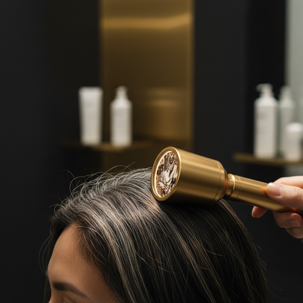
Quick answer: Scalp Dermatoscopy Basics refers to the non-invasive clinical technique of examining the scalp and hair shafts under optical magnification, typically between 10x and 70x, using polarised or non-polarised light. Also known as trichoscopy, it allows clinicians to analyse follicular openings, perifollicular skin, microvasculature, and hair shaft morphology in real time. The main limitation is that while it reveals distinct structural patterns, it cannot replace histological biopsy when diagnosing complex cicatricial alopecias or atypical inflammatory scalp disorders.
Key takeaways
- Trichoscopy magnifies the scalp surface and hair shafts up to 70 times, revealing structures invisible to the naked eye.
- It differentiates between non-scarring conditions like androgenetic alopecia and telogen effluvium without invasive procedures.
- Follicular patterns such as yellow dots, black dots, and white dots indicate specific pathological processes.
- Hair shaft diameter diversity of more than 20 percent is the hallmark diagnostic sign of androgenetic alopecia.
- The technique guides targeted biopsies, reducing patient discomfort and increasing diagnostic accuracy.
Scalp Dermatoscopy Basics: what the evidence shows
Scalp dermatoscopy, formally termed trichoscopy, bridges the gap between gross physical examination and microscopic histology. The technique was standardised in the mid-2000s when dermatologists recognised that polarised handheld dermatoscope lenses could evaluate hair disorders without liquid immersion or skin preparation.
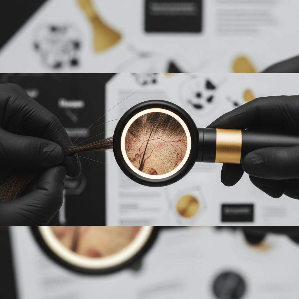
Peer-reviewed studies published in the Journal of the American Academy of Dermatology and the British Journal of Dermatology show that trichoscopy dramatically increases diagnostic precision. In androgenetic alopecia (pattern hair loss), early follicular miniaturisation often escapes naked-eye inspection. Under dermatoscopy, clinicians observe hair shaft diameter diversity exceeding 20 percent across a single field of view. Healthy scalps rarely exceed 10 to 15 percent variability.
Furthermore, trichoscopy allows practitioners to monitor treatment progress objectively. By tracking follicular unit density (the number of single, double, triple, and quadruple hair groupings per square centimetre), clinicians evaluate whether a scalp regimen or medical intervention is preserving follicular output over 6 to 12 month intervals.
The diagnostic mechanism: what the lens reveals
Dermatoscopes illuminate the scalp using cross-polarised light, which cancels surface reflections from the stratum corneum and illuminates deeper sub-epidermal structures. Non-polarised contact dermatoscopy, conversely, provides sharp resolution of the superficial epidermal surface and follicular openings when used with an interface liquid.
During an assessment, clinicians systematically evaluate four distinct anatomical features:
- Follicular openings: The presence, absence, or alteration of the follicular ostia. Empty openings, plugged follicles, or fibrotic scars immediately alter the diagnostic pathway.
- Hair shafts: The calibre, shape, pigmentation, and fracture points of the emerging fibres.
- Perifollicular skin: The colour, scaling, and presence of erythema (redness) around the base of the hair follicle.
- Microvascular structures: The capillary architecture surrounding the follicular units, such as simple loops, twisted loops, or arborising vessels.
Diagnostic signs to look for under magnification
+---------------------+---------------------------------------+---------------------------------------+
| Feature | Characteristic Appearance | Common Clinical Associations |
+---------------------+---------------------------------------+---------------------------------------+
| Yellow Dots | Round, yellowish amorphous structures | Alopecia areata, advanced AGA |
| Black Dots | Dark spots inside empty ostia | Alopecia areata, tinea capitis |
| Exclamation Marks | Hairs thin at base, wide at tip | Active alopecia areata |
| Hair Diversity >20% | Varied hair shaft thicknesses | Androgenetic alopecia (AGA) |
| Peripilar Casts | White or brown rings hugging shaft | Lichen planopilaris, traction |
| White Dots | Fibrotic, shiny white patches | Scarring (cicatricial) alopecia |
| Arborising Red Lines| Branching, enlarged blood vessels | Scalp psoriasis, discoid lupus |
+---------------------+---------------------------------------+---------------------------------------+
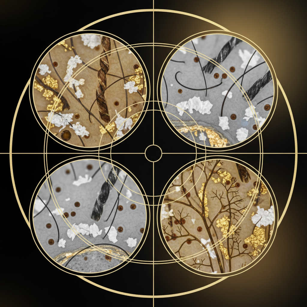
Follicular dots
Dots represent the state of the follicular infundibulum:
- Yellow dots: These are follicular openings distended with sebum and keratinous debris. While typical in severe androgenetic alopecia, large numbers of uniform yellow dots are a classic marker for alopecia areata.
- Black dots: Also known as cadaverised hairs, these represent hair shafts fractured right at the scalp level before emergence. They reflect intense, acute follicular damage, commonly seen in active alopecia areata or tinea capitis (fungal infection).
- White dots: These can be regular and small (normal eccrine duct openings on dark skin) or irregular and large. Large, fibrotic white dots represent destroyed follicles replaced by collagen scar tissue, indicating primary cicatricial alopecia.
Hair shaft abnormalities
Magnification exposes structural defects that differentiate hair shedding from hair breakage:
- Exclamation mark hairs: These hairs taper toward the scalp, demonstrating a damaged, weak root during anagen arrest. They confirm active, expanding alopecia areata.
- Monilethrix and pili torti: Beaded or twisted hair fibres, frequently congenital or caused by severe mechanical and chemical trauma.
- Flame hairs: Wavy, charred-looking remnants that occur when a hair is pulled out forcefully by trichotillomania.
What the evidence does not show
While scalp dermatoscopy provides immense diagnostic clarity, controlled research does not support its use as an absolute standalone test for every condition.
First, dermatoscopy cannot confirm systemic metabolic or nutritional triggers. It cannot detect iron deficiency, thyroid dysfunction, or zinc imbalances. A patient presenting with diffuse hair thinning may show non-specific findings consistent with telogen effluvium, but blood pathology remains mandatory to identify the root driver.
Second, dermatoscopy cannot replace histopathology when distinguishing borderline cases of scarring alopecia, such as early-stage frontal fibrosing alopecia versus severe lichen planopilaris. In such cases, horizontal and vertical section biopsies remain the gold standard.
Third, a dermatoscope is a diagnostic tool, not a therapeutic device. The lens does not stimulate hair follicles, clear sebum plugs, or restore thinning hair. Claims that visualising the scalp under a retail USB microscope can guide miraculous bespoke cures are commercial marketing rather than clinical science.
What you can do: maintaining a healthy scalp environment
Dermatoscopy highlights the critical role of the scalp barrier. Magnified views frequently reveal micro-inflammation, heavy sebum accumulation, and flaky scaling that suffocate the follicular rim long before macroscopic symptoms appear.
To support optimal scalp hygiene:
- Wash regularly: Daily or alternate-day cleansing prevents the accumulation of sebum and oxidised squalene, which appear as yellowish follicular halos under magnification.
- Protect the acid mantle: Use cleansers formulated within a physiological pH range of 4.5 to 5.5. Disrupted surface pH encourages the overgrowth of Malassezia yeasts, triggering inflammatory erythema visible on trichoscopy.
- Avoid prolonged mechanical tension: Trichoscopy readily reveals "hair cast" signs and perifollicular erythema caused by tight ponytails, braids, and extensions, allowing you to loosen hairstyles before permanent traction alopecia occurs.
Supporting scalp wellness with Watermans
When trichoscopy reveals a sluggish scalp environment characterised by product build-up or general dullness without underlying disease, cosmetic support can help maintain optimal conditions. Watermans GrowMe Shampoo and ConditionMe Conditioner are formulated to thoroughly cleanse the follicular environment without stripping natural lipids. Enriched with ingredients like rosemary, biotin, caffeine, and niacinamide, they provide a revitalised, clean canvas that supports the appearance of fuller, thicker, and stronger-feeling hair.
When to get professional medical advice
Do not attempt self-diagnosis using consumer digital scopes. Consult a dermatologist or qualified trichologist if you notice:
- Rapid, patchy hair loss appearing over days or weeks.
- Persistent itching, burning, pain, or visible scaling on the scalp.
- Areas where follicular pores appear completely absent, smooth, or porcelain-like.
- Hair fibres snapping easily along the shaft during routine washing.
Early clinical dermatoscopy can identify cicatricial (scarring) processes before permanent follicular destruction takes place.
Frequently asked questions about Scalp Dermatoscopy Basics
How long does a scalp dermatoscopy examination take?
A routine examination typically takes between 10 and 20 minutes. The clinician systematically surveys multiple anatomical regions including the frontal hairline, vertex, temporal areas, and occiput. They may capture digital images at specific points to establish baseline follicular counts and compare measurements during follow-up visits.
Does a trichoscopy examination hurt?
No, the procedure is completely painless and non-invasive. The clinician gently rests the dermatoscope lens against the scalp surface, parting the hair with a comb. Unlike older diagnostic methods like the surgical hair pluck (trichogram), dermatoscopy does not require pulling hairs out by their roots.
What are the core Scalp Dermatoscopy Basics every clinician looks for?
Every clinician focuses on four primary parameters during an assessment: follicular opening morphology, hair shaft diameter variability, perifollicular skin integrity, and microvascular capillary patterns. Combining these observations allows the practitioner to categorise hair issues accurately.
Can scalp dermatoscopy detect genetic thinning early?
Yes. Scalp dermatoscopy can identify androgenetic alopecia before noticeable thinning occurs. It detects hair shaft diameter diversity exceeding 20 percent and an increase in single-hair follicular units long before diffuse density drops to the point of naked-eye detection.
Can dermatoscopy differentiate hair breakage from genuine hair loss?
Yes. Dermatoscopy instantly clarifies whether hair is snapping along the shaft due to mechanical or chemical trauma, or shedding directly from the follicle. Breakage displays fractured ends (trichorrhexis nodosa) with intact roots, whereas follicular loss displays empty or miniaturised follicular ostia.
Master the essentials of trichoscopy
Understanding Scalp Dermatoscopy Basics transforms how scalp health is evaluated. By making microscopic follicular patterns, vascular changes, and hair shaft structures visible, dermatoscopy removes guesswork from trichological evaluations. While it cannot replace formal lab tests or biopsies in complex diseases, this non-invasive tool provides the clearest, most accessible window into follicular health available in modern dermatology.
Sources and references
- British Association of Dermatologists: Guidelines for the management of alopecia areata (2012).
- Rudnicka, L., et al.: Atlas of Trichoscopy: Dermoscopy in Hair and Scalp Disease, Springer, 2012.
- Journal of the American Academy of Dermatology: Trichoscopy of common hair loss disorders (2008), DOI: 10.1016/j.jaad.2008.06.012.
- International Journal of Trichology: Trichoscopy in hair disorders: an update (2021).
- NHS: Hair loss diagnosis and management guidance (2023).
Find these ingredients in Watermans products
- Zinc: you will find this ingredient in our Hair Vitamins.
- Niacinamide: you will find this ingredient in our Hair Mask.