Follicular Miniaturisation in Androgenetic Alopecia: Biological Mechanisms and Management
9 min read ·
Follicular Miniaturisation in Androgenetic Alopecia causes hair follicles to shrink over time under androgenic influence. Discover the mechanisms and clinical management.
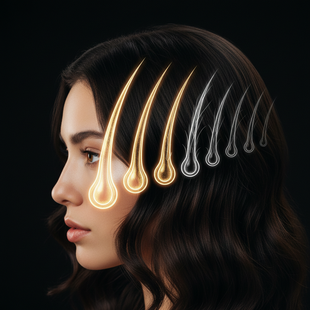
Quick answer: Follicular Miniaturisation in Androgenetic Alopecia is the progressive biological process where genetically predisposed hair follicles shrink under the influence of androgens, specifically dihydrotestosterone (DHT). With each consecutive hair cycle, the growth phase shortens and the dermal papilla decreases in volume, transforming robust terminal hairs into fine, unpigmented vellus-like hairs. While medical interventions can halt progression or partially reverse early shrinkage, long-standing miniaturisation eventually leads to follicular dormancy and fibrosis, making early detection and clinical management vital.
Key takeaways
- Follicular miniaturisation is the central pathological hallmark of pattern hair loss in both men and women.
- The primary driver is local dihydrotestosterone (DHT) binding to androgen receptors in genetically sensitive dermal papillae.
- Each cycle shortens the anagen (growth) phase from several years to just months, while prolonging the telogen (resting) or kenogen (latent) phase.
- Miniaturised hairs remain physically present at first, but their shaft diameter shrinks from over 60 micrometres down to under 30 micrometres.
- Once a follicle undergoes complete fibrosis and loses its stem cell reservoir, miniaturisation becomes irreversible.
Follicular Miniaturisation in Androgenetic Alopecia: what the evidence shows
Follicular Miniaturisation in Androgenetic Alopecia represents a structural downscaling rather than an immediate loss of the follicle. Clinical trichology and dermatopathology demonstrate that hair follicles do not vanish overnight when pattern loss begins. Instead, susceptible follicles undergo an orchestrated regression across repeated hair growth cycles.
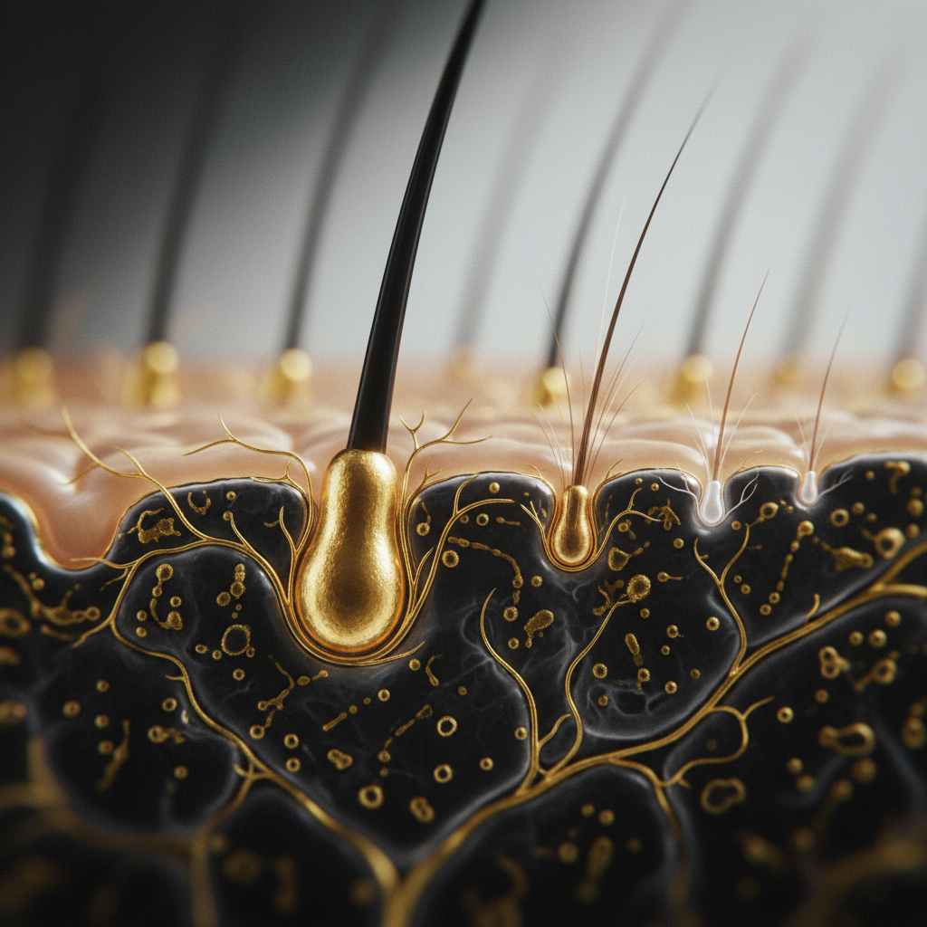
In a healthy scalp, roughly 85 to 90 percent of hairs sit in the anagen growth phase at any given time, where they produce thick, pigmented terminal hair fibres with a shaft diameter between 60 and 80 micrometres. In individuals with androgenetic alopecia, androgen-sensitive follicles in the frontal, vertex, and crown zones respond to circulating and local androgens by truncating this growth window.
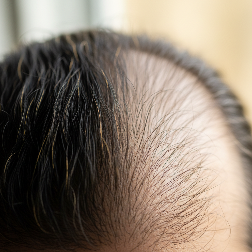
Biopsy studies show that as the follicle shrinks, the dermal papilla loses cells and volume. Consequently, the newly formed hair shaft emerges thinner, shorter, and with reduced melanin content. Dermatologists using dermoscopy (trichoscopy) identify this process by measuring hair diameter diversity: when more than 20 percent of hairs in an affected zone show significant calibre variation (anisotrichosis), active miniaturisation is underway. Over time, terminal hairs transition into vellus-like hairs measuring less than 30 micrometres in diameter, creating the visible appearance of diffuse thinning or receding margins.
The biological mechanism behind miniaturisation
The driving mechanism of follicular miniaturisation relies on an interplay between genetic susceptibility, endocrine signalling, and tissue microenvironments. Testosterone converts to dihydrotestosterone (DHT) via the intracellular enzyme 5-alpha reductase (primarily the Type II isoenzyme located within the dermal papilla and outer root sheath). DHT binds to androgen receptors with approximately five times the affinity of testosterone.
Once DHT binds to these receptors in genetically programmed follicles, it initiates a signalling cascade that alters gene transcription:
- Inhibitory growth factors: The dermal papilla releases transforming growth factor beta (TGF-beta1 and TGF-beta2) and Dickkopf-related protein 1 (DKK-1), which suppress keratinocyte proliferation and prematurely force the follicle out of anagen into catagen (regression).
- Anagen phase truncation: Normal anagen durations of 2 to 7 years shrink progressively down to 1 year, 6 months, or merely weeks.
- Reduction of dermal papilla volume: Fibroblast counts within the dermal papilla decline, directly diminishing the size of the hair bulb and limiting the diameter of the hair fibre it can manufacture.
- Micro-inflammation and fibrosis: Perifollicular lymphocytic infiltrates often cluster around the follicular infundibulum and bulge. Over successive cycles, chronic low-grade inflammation promotes collagen deposition, replacing the follicular tract with fibrous tissue bands (streamers), which permanently prevents the follicle from regenerating a terminal hair.
Terminal hair versus miniaturised hair
To understand the physical impact of miniaturisation, examining how the fibre changes across biological metrics helps clarify why thinning appears long before actual baldness occurs.
| Characteristic | Healthy Terminal Hair | Early Miniaturised Hair | Advanced Miniaturised (Vellus-Like) |
|---|---|---|---|
| Shaft Diameter | 60 to 80 µm | 30 to 55 µm | Under 30 µm |
| Anagen Duration | 2 to 7 years | 1 to 2 years | Several weeks to months |
| Bulb Depth | Deep subcutaneous fat | Reticular dermis | Papillary dermis |
| Pigmentation | Fully pigmented | Reduced pigment | Minimal to no pigment |
| Sebaceous Gland | Normal ratio to follicle | Gland remains intact | Gland appears hypertrophic |
| Reversibility | Baseline healthy state | High potential to recover | Poor / Fibrosed |
What the evidence does not show
Misconceptions around miniaturisation are common in commercial marketing and social discussions. Controlled scientific trials have disproven several widespread assumptions:
- It is not caused by poor scalp blood flow: While local microvascularisation declines alongside the shrinking follicle, this is an effect of the reduced metabolic demand of a smaller organ, not the primary cause. Blood vessels downscale because the tissue requires less perfusion.
- It is not caused by clogged sebum: Sebum production increases visually because the sebaceous gland maintains its original size while the hair follicle below it shrinks. Sebum itself does not obstruct the follicular canal to trigger miniaturisation.
- It is not simple shedding: Telogen effluvium involves full-thickness terminal hairs falling out in high volumes due to systemic stress. Miniaturisation is a gradual change in hair diameter and length, not an abrupt shedding event.
- Cosmetic products cannot alter androgen genetics: Shampoos, conditioners, and superficial cosmetic tonics cannot reprogram androgen receptor sensitivity or reverse established follicular fibrosis.
Clinical signs of active miniaturisation
Recognising early follicular miniaturisation provides the best opportunity for clinical intervention. Key clinical markers include:
- Hair diameter diversity (Anisotrichosis): Viewed under magnification, adjacent follicles display noticeably different thicknesses rather than uniform calibres.
- Loss of hair density without excess shedding: The scalp becomes more visible under bright lighting, yet you may not see abnormal numbers of hairs on your brush or in the shower drain.
- Reduced maximum length: The hair in affected zones (such as the temples or crown) stops growing past a certain short length due to the abbreviated anagen phase.
- Yellow dots and peripilar signs: Dermatologists frequently observe peripilar brown halos (indicating early perifollicular inflammation) or yellow dots (distended follicular ostia filled with sebum where fibres have markedly downscaled).
- Texture changes: The overall volume feels softer, lighter, and more flyaway because the cumulative mass of individual hair fibres has dropped.
Management options: medical treatments and supportive care
Managing miniaturisation focuses on two goals: preventing further downscaling and stimulating existing follicles to produce larger diameters.
Evidence-based medical therapies
First-line interventions recognised by dermatological bodies such as the British Association of Dermatologists (BAD) include:
- 5-alpha reductase inhibitors: Oral finasteride and dutasteride inhibit the conversion of testosterone to DHT, reducing systemic and scalp DHT levels to arrest miniaturisation and allow dermal papillae to recover.
- Vasodilators / Potassium channel openers: Topical or low-dose oral minoxidil extends the anagen phase and widens the follicular aperture, increasing hair shaft calibre over 4 to 12 months of consistent use.
- Anti-androgens: In female pattern hair loss, oral spironolactone or cyproterone acetate can counteract androgen action at the receptor level.
Supportive cosmetic care and scalp hygiene
While cosmetic solutions do not cure genetic conditions, maintaining an optimal scalp environment supports general hair health. A balanced scalp with controlled surface sebum and minimal oxidative stress ensures that emerging fibres remain free from superficial irritation.
Watermans focuses on everyday cosmetic support for individuals experiencing thinning hair. Formulations such as Watermans Grow Me Shampoo and Condition Me incorporate ingredients like caffeine, rosemary extract, biotin, and niacinamide. In cosmetic science, topical caffeine and botanical extracts have been studied for their ability to invigorate the scalp surface, improve moisture retention, and provide cosmetic body to fine, thinning hair. When integrated alongside physician-led treatments, quality cosmetic routines help protect delicate, miniaturised strands from mechanical breakage and improve overall manageable volume.
When hair thinning is caused by something else
Not all thinning stems from androgen-mediated miniaturisation. Several other scalp and systemic conditions mimic these visible changes:
- Diffuse Alopecia Areata: An autoimmune attack targeting the hair bulb can cause rapid, generalised thinning that lacks the classic androgen-dependent pattern.
- Chronic Telogen Effluvium: Ongoing nutritional deficits (such as severe ferritin or vitamin D deficiency), thyroid dysfunction, or chronic metabolic stress cause diffuse shedding across the entire scalp without significant anisotrichosis.
- Cicatricial (Scarring) Alopecias: Conditions such as Lichen Planopilaris (LPP) or Frontal Fibrosing Alopecia (FFA) destroy the follicular stem cells permanently through intense inflammation, replacing follicles with scar tissue without the protracted miniaturisation phase seen in pattern loss.
When to seek professional advice
Consult a GP, dermatologist, or qualified trichologist at the earliest notice of progressive thinning or changes in your hairline. Early assessment is critical because treatments can maintain and thicken miniaturising follicles, but they cannot regenerate tissue that has completely fibrosed.
Seek immediate clinical evaluation if you experience scalp pain, burning, redness, rapid patchy hair loss, or scaling, as these symptoms indicate inflammatory or scarring conditions requiring urgent medical management.
Frequently asked questions about Follicular Miniaturisation in Androgenetic Alopecia
What is Follicular Miniaturisation in Androgenetic Alopecia?
Follicular Miniaturisation in Androgenetic Alopecia is the biological process where hair follicles progressively shrink under the influence of dihydrotestosterone (DHT). With each consecutive cycle, the follicle produces shorter, finer hair shafts, eventually converting thick terminal hairs into thin, vellus-like strands.
Can miniaturised hair follicles be restored to normal size?
Yes, partially miniaturised follicles can often be stimulated to produce thicker hair fibres using clinically proven treatments like minoxidil and 5-alpha reductase inhibitors. However, recovery is only possible if the follicle is still viable; once complete fibrosis occurs, reversal is impossible.
How quickly does follicular miniaturisation progress?
Progress varies widely based on genetic predisposition and androgen levels. It typically develops slowly over several years or decades, though aggressive early-onset androgenetic alopecia in young adults can cause noticeable cosmetic thinning within 12 to 24 months.
Does follicular miniaturisation affect the entire scalp?
No. Miniaturisation specifically targets follicles with genetic sensitivity to androgens, predominantly in the temples, frontal hairline, mid-scalp, and crown. Follicles in the occipital region (the back and lower sides of the head) are usually resistant to androgen-mediated shrinking.
How do dermatologists diagnose miniaturisation?
Clinicians use dermoscopy or trichoscopy to view the scalp under high magnification. The primary diagnostic sign is anisotrichosis, meaning marked diversity in hair shaft diameter across the affected zones, accompanied by peripilar signs and a reduced number of hairs per follicular unit.
Summary of managing follicular miniaturisation
Follicular Miniaturisation in Androgenetic Alopecia is an intricate biological process driven by genetics and hormonal signalling. Understanding that miniaturisation represents a gradual shrinking of the hair factory, rather than sudden fibre detachment, clarifies why proactive care yields the best outcomes. By combining medical interventions that mitigate androgenic signalling with dedicated cosmetic scalp care to reduce mechanical breakage, you can successfully protect follicular health and retain hair volume over the long term.
Sources and references
- British Association of Dermatologists. Clinical Guidelines for the Management of Alopecia Areata and Androgenetic Alopecia. 2021.
- Journal of the American Academy of Dermatology. Dermoscopic features of androgenetic alopecia: A systematic review. 2018.
- International Journal of Molecular Sciences. The Role of Androgens, Androgen Receptors, and 5α-Reductase in Androgenetic Alopecia. 2020.
- NHS. Hair loss: Overview and treatments. 2023.
- Dermatologic Clinics. Pathophysiology and genetics of androgenetic alopecia. 2021.
Find these ingredients in Watermans products
- Vitamin D: you will find this ingredient in our Hair Vitamins.
- Niacinamide: you will find this ingredient in our Hair Mask.