Dermoscopy of the Hair and Scalp: Diagnostic Guide
8 min read ·
Dermoscopy of the Hair and Scalp (trichoscopy) is a non-invasive diagnostic method that uses magnification to assess hair loss, follicles, and scalp disorders.
Quick answer: Dermoscopy of the Hair and Scalp, clinically referred to as trichoscopy, is a non-invasive diagnostic method that uses polarised light and optical magnification (typically 10x to 70x) to evaluate hair shafts, follicular openings, and the surrounding scalp skin. It enables practitioners to distinguish between conditions like androgenetic alopecia, alopecia areata, and scarring alopecias rapidly without an initial biopsy. Its main limitation is that it cannot replace histopathology in complex or atypical cases where cellular pathology remains hidden beneath the basement membrane.
Key takeaways
- Dermoscopy of the Hair and Scalp allows direct, magnified visualisation of follicular units, hair shaft morphology, and vascular patterns.
- It differentiates non-scarring hair loss from scarring forms by identifying the presence or loss of follicular ostia (openings).
- Specific diagnostic markers, such as yellow dots, black dots, exclamation mark hairs, and hair diameter diversity, provide rapid diagnostic guidance.
- While it reduces the need for invasive procedures, a scalp biopsy remains the definitive test when trichoscopic patterns are ambiguous or overlap.
- Trichoscopy is also a valuable tool for tracking treatment response over time by measuring hair shaft thickness and follicular density objectively.
Dermoscopy of the Hair and Scalp: what the evidence shows
Dermoscopy of the Hair and Scalp has transformed clinical trichology over the past two decades. Historically, clinicians relied on naked-eye inspection, the hair pull test, and invasive punch biopsies to diagnose complex scalp disorders. By applying dermoscopic principles to scalp skin, dermatologists can now identify micro-structures that are completely invisible to the naked eye.
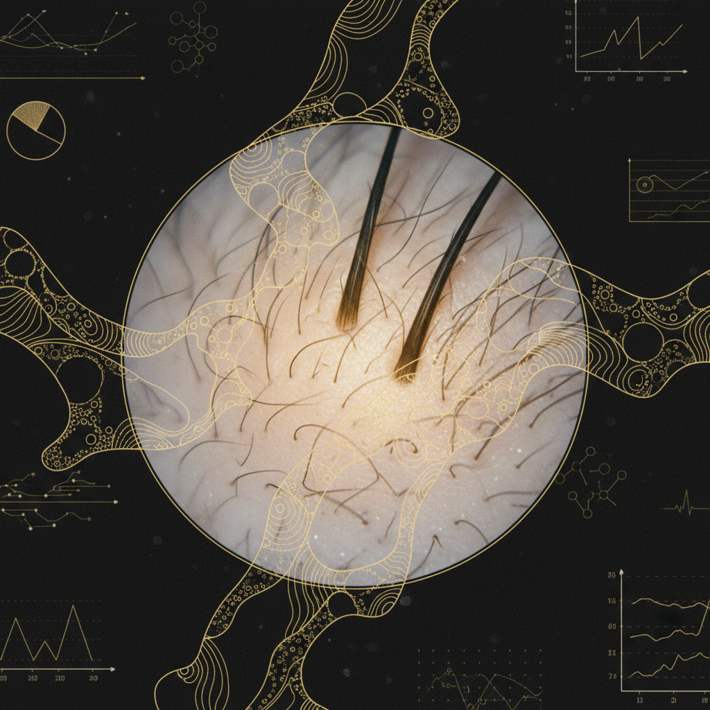
Clinical research published across major dermatology journals demonstrates that trichoscopy dramatically increases diagnostic accuracy. In non-scarring conditions, it reveals distinct follicular and interfollicular features:
- Androgenetic alopecia (pattern hair loss): The hallmark sign is hair diameter diversity greater than 20 percent across the affected area. Clinicians also observe peripilar signs (brown halos around follicular openings indicating micro-inflammation) and an increased proportion of single-hair follicular units compared to triple- or double-hair units.
- Alopecia areata: Active disease is marked by yellow dots (sebum and keratin debris in empty follicles), black dots (cadaverised hairs broken at the scalp surface), micro-exclamation mark hairs (tapered proximal ends), and short vellus hairs during remission.
- Telogen effluvium: Trichoscopy typically shows empty follicular openings and short, regrowing upright hairs without marked hair diameter diversity, helping distinguish it from early androgenetic alopecia.
In primary cicatricial (scarring) alopecias, early detection is vital to prevent irreversible follicular destruction. Dermoscopy of the Hair and Scalp reliably detects the absence of follicular openings, which confirms permanent follicular loss. Conditions such as lichen planopilaris present with prominent peripilar casts (keratotic collars around emerging hair shafts) and perifollicular erythema, while frontal fibrosing alopecia displays lonely hairs along the frontal hairline and pinpoint peripilar scale.
What the evidence does not show
While trichoscopy is a powerful diagnostic aid, it is not an all-seeing oracle. Clinical evidence clearly delineates its diagnostic boundaries:
- It does not replace histopathology in late-stage disease: When inflammatory scarring conditions reach an advanced stage, the scalp often appears as a non-descript, smooth, white plaque devoid of openings. At this point, dermoscopic signs blur, and a 4 mm punch biopsy remains necessary to determine the underlying process.
- It does not identify systemic or nutritional triggers: A trichoscope cannot diagnose iron deficiency, thyroid dysfunction, or hormonal imbalances. It shows the morphological consequences of these issues, not their biochemical root causes. Blood panels are still required.
- It does not provide instantaneous prognosis: Seeing regrowing upright hairs suggests follicular activity, but it cannot guarantee long-term restoration or predict whether an autoimmune flare will recur.
Diagnostic patterns across common scalp conditions
To understand how clinicians read the scalp, it helps to compare the core visual markers across the conditions most frequently encountered in clinical practice.
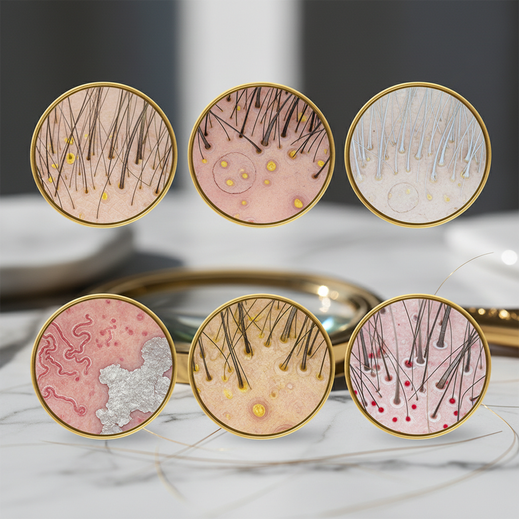
| Condition | Follicular Openings | Hair Shaft Signs | Interfollicular Scalp Skin |
|---|---|---|---|
| Androgenetic Alopecia | Preserved; empty follicles present | Hair diameter diversity (>20%), miniaturised vellus hairs | Peripilar brown halo (inflammation), sun damage in bare areas |
| Alopecia Areata | Preserved; prominent yellow dots | Black dots, exclamation mark hairs, broken hairs | Normal, or mild background erythema |
| Telogen Effluvium | Preserved; normal follicular density | Uniform hair diameter, upright regrowing hairs | Normal, no significant vascular abnormality |
| Lichen Planopilaris | Progressively lost; white patches | Peripilar casts (cylindrical scaling clinging to shaft) | Perifollicular erythema, milky-red areas |
| Discoid Lupus Erythematosus | Lost in fibrotic areas; large keratotic plugs | Dysmorphic hairs, fractured shafts | Thick branching arborising vessels, follicular plugging |
The mechanism: how trichoscopy illuminates the scalp
The biological mechanism of trichoscopy relies on physics and optics. The human stratum corneum reflects roughly 4 to 7 percent of incoming light due to the mismatch in refractive index between air (1.0) and keratinised skin (approximately 1.55). This surface reflection causes glare that obscures deeper epidermis and dermis.
Dermoscopy resolves this in two ways:
- Polarised light: Uses cross-polarised filters to block surface reflections, allowing photons to penetrate down to the superficial dermis. This highlights deeper structures like blood vessels, pigment, and dermal fibrosis.
- Non-polarised light with liquid immersion: Employs an interface fluid (such as mineral oil or alcohol) and a glass contact plate to match the refractive index of the stratum corneum, allowing crystal-clear assessment of superficial scaling and the follicular infundibulum.
By combining these methods, a clinician sees the entire follicular architecture: the infundibulum, the hair shaft thickness, the surrounding vascular network, and the interfollicular skin condition.
Signs to look for: when to investigate further
If you are a salon professional or an individual monitoring your scalp, certain signs visible to the naked eye or under basic magnification indicate that medical dermoscopy of the hair and scalp is required:
- Localised patches devoid of pores: A shiny, porcelain-smooth scalp area where you can no longer see follicle pores indicates potential scarring alopecia.
- Collars of scale surrounding single hairs: Tubular scales that sleeve the base of the hair shaft rather than sitting flat on the scalp.
- Persistent localised redness with burning or itching: Discomfort paired with redness around hair shafts frequently points to active follicular inflammation.
- Abrupt, round bald patches: Clean, circular clearings that appear over a few days or weeks.
- Marked widening of the natural parting: Especially where hairs along the front and crown vary dramatically in thickness.
What you can do: clinical and cosmetic care
When navigating scalp issues, your approach should separate medical treatment from cosmetic support.
Clinical steps
Always seek an evaluation from a dermatologist or qualified trichologist. They will use dermoscopy to map your scalp, identify whether loss is scarring or non-scarring, and measure miniaturisation ratios. If active inflammation or autoimmune activity is present, medical interventions (such as topical anti-inflammatories, intralesional therapies, or systemic treatments) are essential to arrest damage.
Daily cosmetic maintenance
Cosmetic haircare cannot reverse scarring or cure autoimmune disease, but maintaining a clean, balanced scalp environment supports healthy follicular function:
- Cleanse regularly to prevent the accumulation of sebum, dead keratin, and microbial overgrowth around the ostia.
- Avoid mechanical tension that causes traction damage, visible under dermoscopy as empty follicles with perifollicular erythema along tension margins.
- Protect vulnerable scalp areas from ultraviolet radiation. Sun-damaged scalps develop honey-comb pigment networks that complicate dermoscopic tracking.
For individuals experiencing thinning associated with normal shedding and breakage, cosmetic options such as Watermans GrowMe shampoo and conditioners help improve hair fibre condition, strengthen fragile strands, and refresh the scalp skin without interfering with professional dermatological care.
When the cause may be something else
Scalp changes do not always mean a primary alopecia disorder. Several systemic conditions mimic common hair loss patterns:
- Tinea capitis: Fungal infection can produce comma hairs, corkscrew hairs, and zigzag hairs alongside heavy scaling, easily mistaken for alopecia areata or severe seborrhoeic dermatitis.
- Trichotillomania: Compulsive hair pulling creates irregular bald patches featuring hairs broken at uneven lengths, coiled hairs, and flame-shaped hair remnants.
- Systemic lupus or secondary syphilis: Both can manifest as diffuse scalp thinning or "moth-eaten" alopecia that requires serological testing rather than topical treatments alone.
Frequently asked questions about Dermoscopy of the Hair and Scalp
What happens during a dermoscopy of the hair and scalp examination?
A clinician gently parts your hair in multiple zones (frontal, crown, temples, and occiput) and places a handheld dermatoscope against the scalp. The procedure is entirely painless, non-invasive, and takes between 10 and 20 minutes. High-resolution images are often recorded digitally so your hair density, shaft calibre, and follicular health can be tracked precisely over subsequent visits.
Can dermoscopy tell the difference between breakage and hair loss?
Yes. Dermoscopy of the Hair and Scalp easily distinguishes shaft breakage from genuine follicular loss. Broken hairs show irregular, fractured distal ends (trichorrhexis nodosa) with intact roots sitting in the follicle. In contrast, genuine shedding or alopecia shows empty follicular openings, vellus transformation, or tapered roots emerging from the ostium.
Does dermoscopy hurt or require shaving the head?
No. The examination is completely comfortable and does not require shaving or cutting any hair. The practitioner simply uses a fine-tooth comb or probe to part your strands and view the scalp skin directly. If an immersion fluid is used, a small drop of alcohol or oil may feel slightly cool on your skin.
Can a trichoscopy exam replace a scalp biopsy?
In many routine cases of male or female pattern hair loss and classic alopecia areata, trichoscopy provides sufficient diagnostic certainty to avoid a scalp biopsy. However, if a scarring alopecia is suspected or features appear mixed, a small punch biopsy remains the gold standard to assess deep dermal and subcutaneous inflammation.
Dermoscopy of the Hair and Scalp in clinical practice
Dermoscopy of the Hair and Scalp bridges the gap between simple naked-eye observation and invasive surgical biopsies. By providing a clear, magnified window into the follicular unit and surrounding microvasculature, it empowers clinicians to identify alopecias earlier, intervene before scarring causes permanent loss, and measure response to therapies objectively. Whether distinguishing between temporary shedding and progressive thinning or catching an aggressive inflammatory condition in its infancy, this non-invasive tool remains one of modern trichology's most vital clinical assets.
Sources and references
- British Association of Dermatologists. Guidelines for the management of alopecia areata. 2012.
- Rudnicka, L., Olszewska, M., Rakowska, A., & Slowinska, M. Atlas of Trichoscopy: Dermoscopy in Hair and Scalp Disease. Springer, 2012.
- Miteva, M., & Tosti, A. "Hair and scalp dermatoscopy." Journal of the American Academy of Dermatology, 2012. DOI: 10.1016/j.jaad.2012.02.013.
- Rakowska, A. "Trichoscopy (hair and scalp videodermoscopy) in the healthy female. Method standardization and acceptable norms." Journal of Dermatological Case Reports, 2009. DOI: 10.3315/jdcr.2009.1028.
- National Institute for Health and Care Excellence (NICE). Alopecia: clinical management overview. 2023.