Contact Dermatitis On Scalp: Causes, Diagnosis, and Barrier Recovery
9 min read ·
An evidence-based guide to Contact Dermatitis On Scalp: learn the differences between irritant and allergic reactions, clinical symptoms, and clear recovery steps.
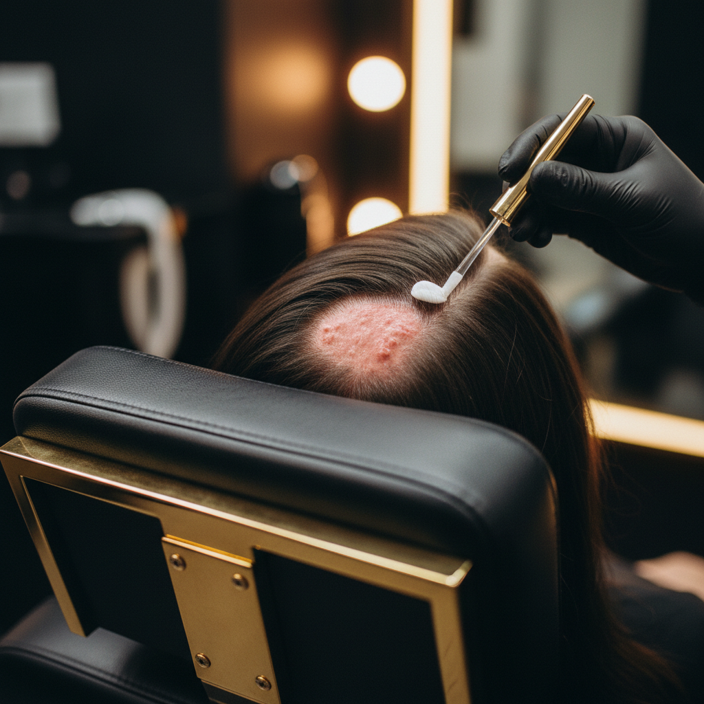
Quick answer: Contact Dermatitis On Scalp is an inflammatory skin reaction triggered either by direct irritation from harsh substances or by an allergic response to specific topical ingredients. It typically presents with intense itching, redness, burning, tenderness, and scaling along the hair-bearing skin and hairline. Identifying and eliminating the offending chemical is the definitive treatment, though severe or persistent reactions require clinical diagnosis and targeted topical medical therapy to resolve safely without causing secondary skin complications.
Key takeaways
- Scalp contact dermatitis occurs in two forms: irritant contact dermatitis (non-immune damage to the skin barrier) and allergic contact dermatitis (a type IV delayed hypersensitivity response).
- Common culprits include paraphenylenediamine (PPD) in permanent hair dyes, fragrances, preservatives like methylisothiazolinone, and harsh surfactants.
- Symptoms often extend beyond the crown to the hairline, forehead, neck, and ears, where the skin is thinner and more permeable.
- While acute dermatitis causes temporary hair shedding (telogen effluvium) through scalp inflammation, it does not directly cause permanent follicular scarring unless secondary infection occurs.
- Patch testing performed by a dermatologist remains the clinical gold standard for distinguishing allergic reactions from other scalp conditions.
Contact Dermatitis On Scalp: what the evidence shows
Contact Dermatitis On Scalp represents a frequent diagnostic challenge in trichology and clinical dermatology. Clinical surveillance data from the British Association of Dermatologists and international contact dermatitis research groups consistently identify haircare products as major drivers of head and neck dermatitis.
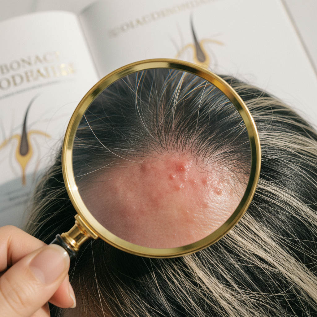
The scalp possesses a robust epidermal barrier compared to facial skin, heavily populated by sebaceous glands and terminal hair follicles. Because of this thicker stratum corneum, the hair-bearing scalp can sometimes tolerate strong irritants better than surrounding zones. Consequently, a reaction caused by a shampoo, bleach, or dye may manifest primarily on the upper neck, eyelids, behind the ears, or along the hairline before obvious signs emerge across the crown itself.
Evidence demonstrates that contact dermatitis falls into two primary categories:
- Irritant Contact Dermatitis (ICD): This accounts for approximately 80 percent of all contact dermatitis cases. It is a non-immunological reaction resulting from direct physical or chemical damage to keratinocytes and the intercellular lipid matrix. High-pH alkaline relaxers, concentrated hydrogen peroxide (bleach), and aggressive anionic surfactants can strip the lipid barrier rapidly, producing immediate burning, stinging, and erythema.
- Allergic Contact Dermatitis (ACD): This represents a cell-mediated, type IV delayed hypersensitivity immune reaction. Sensitisation occurs on initial or repeated exposure. Once primed, T-lymphocytes mount an inflammatory response 24 to 72 hours after re-exposure. Paraphenylenediamine (PPD), botanical extracts, nickel from styling tools, and preservatives such as methylchloroisothiazolinone (MCI) are frequent triggers.
The following table contrasts the two forms to assist with practical differentiation:
| Feature | Irritant Contact Dermatitis (ICD) | Allergic Contact Dermatitis (ACD) |
|---|---|---|
| Primary Mechanism | Non-immune physical or chemical barrier disruption | Type IV delayed hypersensitivity immune reaction |
| Onset of Symptoms | Often immediate or within minutes to hours of application | Delayed, typically appearing 24 to 72 hours post-contact |
| Typical Sensations | Burning, stinging, acute pain, tight sensation | Intense itching, pricking, soreness |
| Physical Signs | Dryness, fissures, superficial scaling, sharp margins | Spreading redness, oedema, vesicles, oozing, crusting |
| Common Triggers | Bleach, chemical straighteners, harsh surfactants, ethanol | PPD (hair dyes), fragrances, MCI/MI preservatives, cocamidopropyl betaine |
| Diagnostic Test | Clinical diagnosis based on exposure history | Epicutaneous patch testing |
What the evidence does not show
Research does not show that Contact Dermatitis On Scalp permanently destroys healthy hair follicles under ordinary conditions. While acute inflammation can disrupt the hair cycle and trigger a temporary shedding phase (telogen effluvium) two to three months later, it does not induce androgenetic alopecia or cicatricial alopecia unless severe chemical burns or untreated bacterial superinfections produce deep dermal scarring.
Furthermore, evidence does not support the popular assumption that "natural", "organic", or "botanical" haircare products are inherently hypoallergenic. Many essential oils, plant extracts (such as chamomile, tea tree oil, and lavender), and natural resins contain potent sensitising compounds capable of eliciting severe allergic contact dermatitis on sensitive scalps.
The biological mechanisms behind the reaction
The development of scalp contact dermatitis hinges on barrier disruption and cellular signalling pathways.
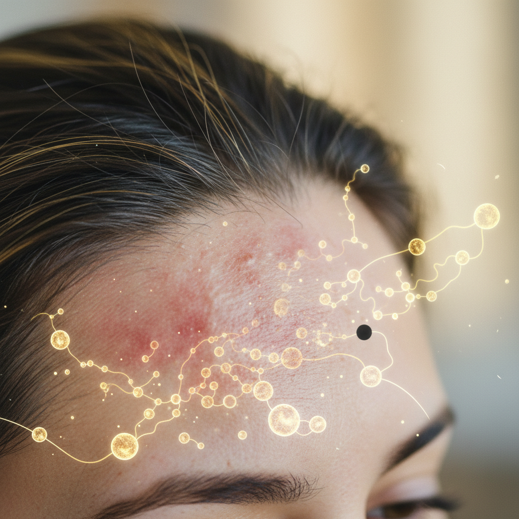
In irritant dermatitis, chemical agents dissolve the stratum corneum's intercellular lipids (ceramides, cholesterol, and free fatty acids). This increases transepidermal water loss (TEWL) and allows direct penetration of the chemical into deeper epidermal layers. The damaged keratinocytes release preformed pro-inflammatory cytokines, specifically interleukin-1 alpha (IL-1α) and tumour necrosis factor-alpha (TNF-α). These signalling proteins recruit neutrophils and macrophages, causing rapid local inflammation, microvascular dilation, and visible redness.
In allergic contact dermatitis, the offending chemical acts as a hapten: a low-molecular-weight molecule that penetrates the stratum corneum and binds to endogenous skin proteins. Epidermal dendritic cells (Langerhans cells) internalise these hapten-protein conjugates, migrate to regional lymph nodes, and present them to naive T-lymphocytes. This primes the immune system. Upon subsequent re-exposure, memory T-cells migrate back to the scalp, releasing interferon-gamma (IFN-γ) and interleukins that trigger spongiosis (intercellular fluid accumulation in the epidermis), vesicle formation, and profound pruritus.
Signs and symptoms to look for
The clinical picture of contact dermatitis can vary from subtle dryness to dramatic swelling. Common manifestations include:
- Pruritus and dysaesthesia: Persistent itching, burning, tingling, or tenderness across the crown, nape, or temples.
- Erythema: Diffuse or patchy redness, frequently spilling over onto the forehead, retroauricular folds (behind the ears), and upper neck.
- Epidermal scaling and flaking: Fine, powdery desquamation or thick, adherent yellowish flakes that may be mistaken for severe dandruff.
- Vesiculation and weeping: In acute allergic episodes, tiny fluid-filled blisters (vesicles) can form, rupture, and weep serous fluid, drying into honey-coloured crusts.
- Facial and periorbital oedema: Severe reactions to hair dyes containing PPD frequently produce significant swelling of the eyelids, forehead, and ears.
When the cause may be something else
Distinguishing contact dermatitis from other inflammatory scalp dermatoses is crucial for appropriate management:
- Seborrhoeic dermatitis: Characterised by greasy, yellowish scales over an erythematous base, primarily targeting areas rich in sebaceous glands (scalp, eyebrows, nasolabial folds). It is driven by Malassezia yeast proliferation rather than an exogenous contact allergen.
- Scalp psoriasis: Typically presents with sharply demarcated, salmon-pink plaques covered with thick, silvery-white micaceous scales, often extending beyond the hairline into the forehead. Auspitz sign (pinpoint bleeding on scale removal) is often positive.
- Atopic dermatitis: Part of a broader systemic atopic diathesis, usually presenting with flexural eczema and a personal or family history of asthma or allergic rhinitis.
- Folliculitis: Discrete pustules and papules centred strictly on hair follicles, driven by bacterial (e.g. Staphylococcus aureus) or fungal colonisation, rather than diffuse epidermal erythema.
Practical steps to take
If you suspect an acute reaction to a hair product, structured management reduces inflammation and protects the skin barrier:
- Immediate cessation: Stop using all suspected products immediately. This includes recent colourants, toners, styling waxes, leave-in serums, and essential oils.
- Thorough rinse: Gently cleanse the hair and scalp with tepid water (avoid hot water, which increases capillary dilation and itching) to dilute and remove any residual chemical residue.
- Simplify the regimen: Strip your routine down to a minimal, fragrance-free, neutral-pH cleanser designed for sensitive skin. Avoid aggressive physical scrubbing, scalp brushes, and thermal styling tools while the skin heals.
- Cool compresses: Applying clean, damp towels chilled with cool water to the scalp for 10 to 15 minutes can soothe burning sensations and reduce acute swelling.
- Seek patch testing: For recurrent or unresolving reactions, consult a dermatologist for standardised epicutaneous patch testing (such as the European Baseline Series) to identify the precise chemical culprit.
Supporting scalp recovery with gentle care
During and immediately following a flare of Contact Dermatitis On Scalp, the skin requires gentle support to restore its acid mantle and replenish depleted lipids. Formulations that avoid common contact allergens, drying alcohols, and aggressive sulfates help the barrier recover without provoking further inflammation.
Watermans products are formulated with an emphasis on scalp health, maintaining an optimal pH balance to support a calm, clean environment without disrupting delicate skin barrier structures. Incorporating nourishing, targeted haircare into a stable routine helps reinforce hair fibre resilience against mechanical stress while the underlying scalp tissue regains physiological stability.
When to seek medical advice
You should consult a GP or dermatologist immediately if you experience:
- Rapid swelling spreading to the eyelids, lips, face, or neck.
- Difficulty breathing or swallowing, which indicates a medical emergency (anaphylaxis).
- Extreme pain, hot skin, yellow purulent discharge, or red streaking, indicating secondary bacterial infection (impetiginisation or cellulitis).
- Blisters covering large portions of the scalp.
- Symptoms that fail to improve within one week of discontinuing suspected products.
Frequently asked questions about Contact Dermatitis On Scalp
How long does Contact Dermatitis On Scalp take to clear up?
Once the offending trigger is completely removed, mild irritant contact dermatitis generally resolves within 3 to 7 days as the epidermal barrier repairs itself. Allergic contact dermatitis takes longer to clear, typically requiring 2 to 3 weeks for activated immune cells to decline and the stratum corneum to fully regenerate. Severe episodes with weeping or crusted skin may need short-term prescription topical corticosteroids to clear promptly.
Can hair dye cause sudden allergic contact dermatitis after years of safe use?
Yes. Sensitisation to allergens like paraphenylenediamine (PPD) is acquired over time through repeated exposure. You can use a permanent hair dye for years without issue until the immune system crosses a threshold and becomes sensitised. Once sensitisation occurs, subsequent contact triggers an acute allergic reaction within 24 to 72 hours, which is why pre-colour patch tests remain essential before every single application.
Does contact dermatitis on the scalp cause permanent hair loss?
No, contact dermatitis does not damage the underlying hair follicles or induce permanent follicular scarring under normal circumstances. However, intense local inflammation or chronic, aggressive scratching can cause temporary hair shedding (telogen effluvium) or mechanical hair breakage along the shaft. The hair regrowth cycle returns to normal once the scalp inflammation resolves and the skin barrier recovers.
What is the difference between contact dermatitis and dandruff?
Dandruff is a non-inflammatory or minimally inflammatory form of seborrhoeic dermatitis driven by an overgrowth of Malassezia yeast and natural sebum degradation, leading to oily, white or grey flakes without acute tenderness. Contact dermatitis, by contrast, is an exogenous reaction to an applied chemical, causing acute redness, burning, sharp itching, and sometimes vesicles or weeping along the hairline.
Clear identification supports long-term scalp health
Managing Contact Dermatitis On Scalp successfully requires careful observation, elimination of offending chemicals, and respect for the epidermal barrier. By learning to distinguish between direct irritation and true allergic hypersensitivity, you can make informed product choices, avoid repeat exposures, and maintain a calm, healthy foundation for sustained hair vitality.
Sources and references
- British Association of Dermatologists. Clinical Guidelines for the Management of Contact Dermatitis.
- National Institute for Health and Care Excellence (NICE). Dermatitis - contact: Scenario: Scalp dermatitis.
- European Society of Contact Dermatitis (ESCD). Guidelines for diagnostic patch testing in dermatitis.
- McFadden, J.P. Et al. Hair dye allergy and paraphenylenediamine: clinical review and management. Contact Dermatitis journal.
- American Academy of Dermatology (AAD). Contact Dermatitis: Diagnosis and Treatment.