Scarring Alopecia Types: Understanding Cicatricial Hair Loss
10 min read ·
Learn about the main Scarring Alopecia Types, their biological mechanisms, symptoms, and why early diagnosis is vital to prevent permanent hair loss.
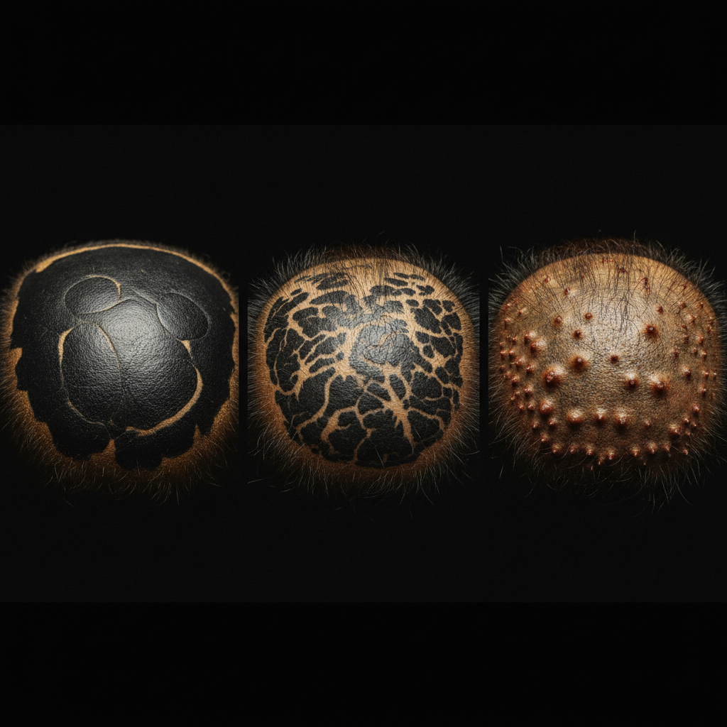
Quick answer: Scarring Alopecia Types refer to a diverse group of rare inflammatory scalp disorders, scientifically termed cicatricial alopecias, where immune cells destroy hair follicles and replace them with fibrous scar tissue. This destruction causes irreversible, permanent hair loss. Clinicians classify these conditions primarily by the dominant immune cells involved: lymphocytic, neutrophilic, or mixed. The crucial limitation is that destroyed follicles cannot regrow; medical treatment focuses entirely on halting active inflammation to protect surviving hair follicles before permanent scarring takes hold.
Key takeaways
- Cicatricial conditions permanently destroy the hair follicle stem cell niche located in the follicular bulge, replacing healthy tissue with collagen scar tissue.
- The North American Hair Research Society classifies primary scarring alopecias into three main cellular categories: lymphocytic, neutrophilic, and mixed.
- Common lymphocytic forms include Lichen Planopilaris (LPP), Frontal Fibrosing Alopecia (FFA), and Central Centrifugal Cicatricial Alopecia (CCCA).
- Common neutrophilic forms include Folliculitis Decalvans and Dissecting Cellulitis of the scalp.
- Early diagnosis via dermoscopy and scalp punch biopsy is essential because cosmetic treatments cannot restore fibrosed, destroyed follicles.
- Medical therapy relies on anti-inflammatory agents, corticosteroids, antimalarials, and targeted antibiotics directed by a dermatologist.
Scarring Alopecia Types: what the evidence shows
Medical consensus divides cicatricial alopecia into primary and secondary categories. In secondary scarring alopecia, follicular destruction occurs as collateral damage from an external insult, such as a severe thermal burn, radiation therapy, cutaneous malignancy, or deep mechanical trauma. In primary scarring alopecia, the hair follicle itself is the specific target of an inflammatory, autoimmune, or autoinflammatory assault.
To standardise diagnosis and research, the North American Hair Research Society (NAHRS) established a classification framework based on the histological inflammatory infiltrate seen under microscopic examination. A dermatopathologist identifies the primary white blood cells attacking the tissue, categorising the disorder into one of three distinct groups:
1. Lymphocytic Scarring Alopecia
In this category, T-lymphocytes infiltrate the upper portion of the follicle. The most prevalent forms include:
- Lichen Planopilaris (LPP): Causes scattered, patchy hair loss accompanied by violaceous erythema (purplish redness) and severe perifollicular hyperkeratosis (scaling collars around the hair base). It predominantly affects middle-aged adults.
- Frontal Fibrosing Alopecia (FFA): Considered an LPP variant, FFA presents as a progressive, symmetrical band-like recession along the frontal and temporal hairline, frequently accompanied by the partial or complete loss of eyebrows.
- Central Centrifugal Cicatricial Alopecia (CCCA): Primarily affects women of African descent, initiating at the vertex or crown and radiating outward symmetrically. Follicular damage is accompanied by progressive thinning and tenderness.
- Pseudopelade of Brocq: An end-stage, indolent condition characterised by smooth, porcelain-white patches of hair loss resembling footprints in the snow, often showing minimal visible active inflammation.
2. Neutrophilic Scarring Alopecia
Neutrophils, the body's acute first-responder immune cells, drive severe suppurative (pus-forming) inflammation around the follicle.
- Folliculitis Decalvans (FD): Presents with recurrent pustules, erosions, crusted lesions, and tufted hairs (polytrichia), where multiple hair shafts emerge from a single dilated follicular opening like a doll's hair. Secondary bacterial colonisation with Staphylococcus aureus is common.
- Dissecting Cellulitis of the Scalp (Perifolliculitis Capitis Abscedens et Suffodiens): Part of the follicular occlusion tetrad, this severe condition causes painful, fluctuating nodules, communicating sinuses, and purulent drainage, predominantly in young men of African descent.
3. Mixed Infiltrate Scarring Alopecia
These disorders exhibit an inflammatory profile featuring both lymphocytes and neutrophils, or foreign-body giant cells, as the disease evolves.
- Folliculitis Keloidalis (Acne Keloidalis Nuchae): Typically presents at the nape of the neck or occipital scalp as firm, follicular papules that gradually coalesce into fibrotic, keloid-like plaques.
- Folliculitis (Tufted) and Erosive Pustular Dermatosis: Chronic inflammatory disorders characterised by recurrent crusts, pustules, and superficial erosions, often triggered by actinic damage or skin trauma in older adults.
| Scarring Alopecia Classification | Predominant Cell Infiltrate | Typical Clinical Presentation | Common Diagnoses |
|---|---|---|---|
| Lymphocytic | T-lymphocytes | Perifollicular erythema, scaling collars, band recession | Lichen Planopilaris, Frontal Fibrosing Alopecia, CCCA |
| Neutrophilic | Neutrophils | Pustules, boggy nodules, abscesses, tufted hairs | Folliculitis Decalvans, Dissecting Cellulitis |
| Mixed | Lymphocytes, neutrophils, plasma cells | Firm papules, keloidal scars, chronic crusting | Acne Keloidalis Nuchae, Erosive Pustular Dermatosis |
| Secondary | Variable (non-specific) | Diffuse fibrosis following external tissue destruction | Thermal burns, radiation, severe infection, surgical trauma |
What the evidence does not show
Controlled research has not found that primary scarring alopecia is caused by routine cosmetic habits, everyday hair washing, or poor hygiene. While excessive mechanical tension and chemical relaxers have been studied as potential contributing or exacerbating factors in CCCA and traction-related scarring, clinical evidence shows these practices do not act as the sole cause. A primary genetic, immunological, and structural susceptibility is required.
Furthermore, evidence does not show that non-prescription products, essential oils, or dietary supplements can halt or reverse active scarring alopecia. Because the pathology involves an autoimmune or profound autoinflammatory cascade that destroys follicular stem cells, gentle cosmetic care cannot replace targeted medical intervention.
The biological mechanism of permanent hair loss
To understand why hair loss is irreversible across all Scarring Alopecia Types, you must look at the follicular bulge. Located in the outer root sheath near the insertion site of the arrector pili muscle, the bulge houses the multipotent epithelial stem cells responsible for regenerating the lower hair follicle during each new anagen (growth) cycle.
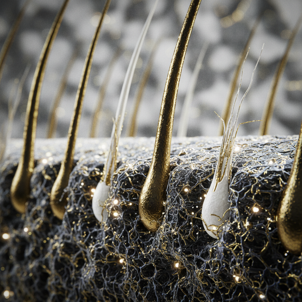
In non-scarring conditions like telogen effluvium or early androgenetic alopecia, inflammation affects the lower bulb or involves microscopic follicular miniaturisation, leaving the stem cells in the bulge unharmed. The follicle retains its regenerative machinery.
In primary cicatricial alopecia, the inflammatory process targets the upper follicle (the infundibulum and isthmus), where immune privilege collapses. Cytotoxic T-cells or reactive neutrophils release destructive cytokines, degrading the basement membrane and triggering apoptosis of the follicular stem cells. Once these cells are destroyed, fibroblasts migrate into the void, laying down dense, unyielding collagen bundles. The sebaceous gland is destroyed early in this process, and the follicular opening vanishes completely, leaving a smooth, atrophic scalp surface devoid of follicular ostia.
Signs and symptoms to watch for
Early identification of Scarring Alopecia Types can prevent widespread loss. While non-scarring shedding presents with diffuse thinning or shedding across the scalp, scarring conditions usually produce focal scalp changes:
- Loss of follicular ostia: Under close examination, the scalp skin appears smooth and shiny, with no visible pores where hairs once emerged.
- Perifollicular erythema and scaling: Redness, violet discolouration, or thick white flakes wrapping around the base of individual hair fibres.
- Subjective sensory symptoms: Persistent burning, tenderness, pruritus (itching), or a crawling sensation in the affected areas.
- Tufted hairs (Polytrichia): Multiple hair shafts (often 5 to 20) clustering out of a single compromised follicular pore.
- Follicular pustules or crusts: Pimples, oozing crusts, or bleeding spots that do not resolve with regular cleansing.
When the cause may be something else
It is vital to distinguish cicatricial alopecia from non-scarring forms of hair loss, as treatments differ radically.
- Androgenetic Alopecia (Pattern Thinning): Produces progressive miniaturisation where thick terminal hairs turn into fine vellus hairs. The follicular openings remain visible, and the scalp exhibits no active erythema, pustules, or scarring.
- Alopecia Areata: An autoimmune attack causing round, smooth patches of hair loss. However, alopecia areata attacks the hair bulb rather than the bulge stem cells. The ostia remain open, exclamation point hairs may appear at margins, and hair can fully regrow.
- Telogen Effluvium: Causes sudden, diffuse shedding across the entire scalp, typically triggered 2 to 3 months after physiological stress or illness. Follicular structures remain intact.
- Traction Alopecia: Initially begins as non-scarring breakage and mechanical loosening from tight hairstyles. If tension continues unresolved over many years, it can progress into secondary cicatricial alopecia with permanently destroyed follicles.
What you can do: medical management and gentle cosmetic care
If you suspect a scarring process, the priority is clinical evaluation. Only a medical professional can arrest the underlying inflammatory destruction.
Medical Treatment
Dermatologists tailor therapy according to the cellular type:
- For lymphocytic disorders: High-potency topical or intralesional corticosteroids, calcineurin inhibitors (tacrolimus), hydroxychloroquine, doxycycline, or systemic immunomodulators to suppress T-cell activation.
- For neutrophilic disorders: Targeted oral antibiotics (such as rifampicin and clindamycin combinations), topical antiseptics, and in severe cases of dissecting cellulitis, oral retinoids or biologic therapies.
Supportive Scalp Care and Watermans
While medical therapies manage the internal inflammatory process, supportive topical care helps maintain the general cosmetic health of the surrounding scalp and surviving hair fibres. Using gentle, non-irritating, sulphate-free cleansers prevents unnecessary mechanical stress on fragile hair.
In this context, Watermans products offer supportive cosmetic conditioning for surviving hair shafts. Formulated with gentle surfactants, biotin, and rosemary, Watermans cleansers and conditioners help cleanse debris from the scalp without stripping delicate moisture. They strengthen the hair fibre against everyday breakage and improve the manageable appearance of existing hair, serving as a cosmetic complement alongside a physician-led dermatological plan.
When to get professional advice
You should consult a GP or a consultant dermatologist promptly if you observe:
- Patches of hair loss that look unusually smooth, shiny, or waxy, with missing pores.
- Unexplained, persistent scalp pain, burning, or intense tenderness.
- Recurrent pustules, boils, or crusted sores on the scalp.
- A rapidly receding frontal hairline accompanied by thinning eyebrows.
A dermatologist will perform dermoscopy (trichoscopy) and usually a 4 mm scalp punch biopsy extending into the subcutaneous fat. This biopsy provides the cellular analysis required to pinpoint the exact condition and begin treatment before scarring spreads.
Frequently asked questions about Scarring Alopecia Types
Can hair regrow once scarring alopecia has occurred?
No. Once a follicle is fully replaced by fibrotic scar tissue, regrowth is biologically impossible because the epithelial stem cells in the follicular bulge are destroyed. Treatment aims to suppress inflammation, relieve discomfort, and protect all remaining, unscarred hair follicles from further permanent damage.
How are different Scarring Alopecia Types accurately diagnosed?
Diagnosis requires clinical trichoscopy and a scalp punch biopsy. A dermatologist takes a small skin sample, typically from the active, inflamed edge of a lesion, and sends it for histopathological evaluation. This reveals whether lymphocytes, neutrophils, or mixed immune cells are driving the condition, confirming the specific diagnosis.
Are primary Scarring Alopecia Types contagious?
No, none of the primary forms are contagious. While secondary bacterial colonisation (such as Staphylococcus aureus) often occurs in neutrophilic conditions like Folliculitis Decalvans, the underlying disease is an inflammatory or autoinflammatory process within the individual's immune system, not an infectious illness that can spread to others.
Is Frontal Fibrosing Alopecia considered one of the Scarring Alopecia Types?
Yes. Frontal Fibrosing Alopecia (FFA) is a primary lymphocytic scarring alopecia. It predominantly affects postmenopausal women, causing progressive hairline recession and eyebrow loss. It is classified clinically and histopathologically as a variant of Lichen Planopilaris.
What should I avoid if I receive a diagnosis of scarring alopecia?
Avoid harsh chemical treatments (such as bleaching or chemical relaxers), excessive heat styling, and tight, pulling hairstyles that add mechanical tension to inflamed follicles. Never delay medical care in favour of unproven cosmetic remedies, as early prescription therapy is the only proven method to halt progression.
Halting the progression of cicatricial hair loss
Understanding the different Scarring Alopecia Types makes one principle clear: early medical detection is the deciding factor in preserving your hair. Whether the destructive infiltrate is lymphocytic, neutrophilic, or mixed, cicatricial alopecia represents a true dermatological emergency for the hair follicle. Cosmetic products and gentle routines cannot rescue stem cells that have already succumbed to fibrosis, but prompt specialist diagnosis, precise biopsy analysis, and targeted anti-inflammatory medical treatment can halt active disease, soothe chronic scalp symptoms, and safeguard your surviving hair for the future.
Sources and references
- North American Hair Research Society (NAHRS), "Classification of Primary Cicatricial Alopecias," Journal of the American Academy of Dermatology, 2001.
- British Association of Dermatologists, "Lichen Planopilaris Clinical Guidelines," BAD Guidelines, 2020.
- National Institute for Health and Care Excellence (NICE), "Alopecia: Summary of Evidence and Management," Clinical Knowledge Summaries, 2022.
- Harries, M. J., et al., "Management of primary cicatricial alopecias: a practical approach," Clinical and Experimental Dermatology, 2018.
- Filbrandt, R., et al., "Primary Cicatricial Alopecia: Diagnosis and Treatment," American Family Physician, 2013.
Find these ingredients in Watermans products
- Biotin: you will find this ingredient in our Hair Growth Shampoo.