Folliculitis Decalvans Overview: Causes, Signs, and Management
8 min read ·
A clinical Folliculitis Decalvans Overview detailing causes, symptoms like tufted hairs, scarring risks, and evidence-based medical treatments to prevent hair loss.
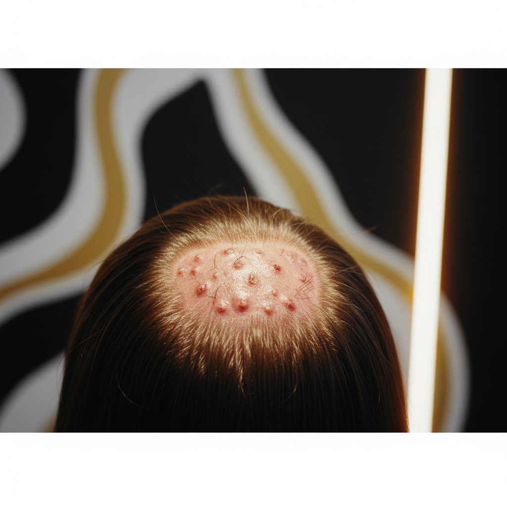
Quick answer: A comprehensive Folliculitis Decalvans Overview reveals that this condition is a rare, chronic primary cicatricial (scarring) alopecia characterised by recurrent follicular pustules, crusting, and progressive, irreversible hair loss. Most commonly driven by a dysregulated immune response to Staphylococcus aureus colonisation, it leads to follicle destruction and tufted hairs. Because destroyed follicles are replaced by fibrotic scar tissue, hair cannot regrow in affected areas. Early medical intervention by a dermatologist is essential to arrest progression and preserve remaining follicles.
Key takeaways
- Folliculitis decalvans is a primary neutrophilic scarring alopecia that causes permanent follicular destruction.
- The hallmark clinical feature is "tufted folliculitis", where multiple hair shafts (often 5 to 20) emerge from a single dilated follicular opening like a doll's hair.
- While Staphylococcus aureus is routinely isolated from active pustules, the root cause involves an abnormal host immune response rather than a simple surface infection.
- Over-the-counter shampoos, hair growth serums, and standard cosmetic treatments cannot reverse scarring or cure the condition.
- First-line medical therapy typically relies on targeted oral antibiotics, often combinations such as rifampicin and clindamycin, to induce remission.
Folliculitis Decalvans Overview: what the evidence shows
In any clinical Folliculitis Decalvans Overview, dermatologists classify the condition as a chronic neutrophilic cicatricial alopecia. It accounts for roughly 10 percent of all primary scarring hair loss cases seen in specialist hair clinics. The condition predominantly affects young to middle-aged adults, with a slight predilection for males, though it can present in anyone regardless of sex or ethnicity.
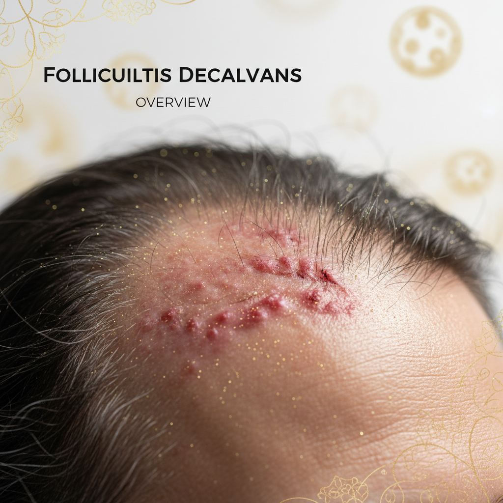
Histopathological studies show that the disease begins with an intense neutrophilic infiltrate around the upper portion of the hair follicle (the infundibulum). As the inflammation advances, it breaches deeper into the follicle, destroying the sebaceous glands and follicular stem cells located in the bulge region. Once these stem cells are destroyed, the follicle cannot undergo normal cycling. The human body replaces the destroyed architectural structure with fibrous connective tissue, culminating in smooth, shiny patches of permanent hair loss.
Bacteriological cultures from active lesions almost universally identify Staphylococcus aureus. However, clinical evidence confirms that folliculitis decalvans is not merely a contagious bacterial infection. Millions of people carry S. Aureus asymptomatically in their nares and on their skin. In patients with folliculitis decalvans, researchers observe an altered cell-mediated immune profile and a heightened inflammatory response to bacterial superantigens. This triggers an uncontrolled cascade of tissue-destructive enzymes, primarily matrix metalloproteinases and pro-inflammatory cytokines.
What the evidence does not show
It is just as vital to understand what folliculitis decalvans is not. Evidence clearly refutes several common misconceptions held by clients and stylists:
- It is not caused by poor hygiene. Folliculitis decalvans is not the result of infrequent washing, dirty styling tools, or oily scalp build-up. Aggressive washing or harsh physical exfoliation does not clear the disease and can exacerbate inflammation.
- It is not contagious. You cannot catch folliculitis decalvans from salon shears, shared towels, pillows, or physical contact with an affected individual.
- It is not a cosmetic condition. While superficial folliculitis can often be managed with gentle antiseptic washes, folliculitis decalvans involves deep follicular architecture. Cosmetic cleansers, essential oils, and topical anti-dandruff solutions cannot reach the dermal depth required to halt the disease.
- Scarred follicles cannot be regenerated. Once a patch becomes fibrotic, no cosmetic product, laser device, or topical stimulant can trigger regrowth. Medical therapies focus solely on stopping active disease spread.
Clinical signs and symptoms to look for
Recognising the early presentation of this condition can make the difference between minor localised thinning and extensive, disfiguring scalp baldness.
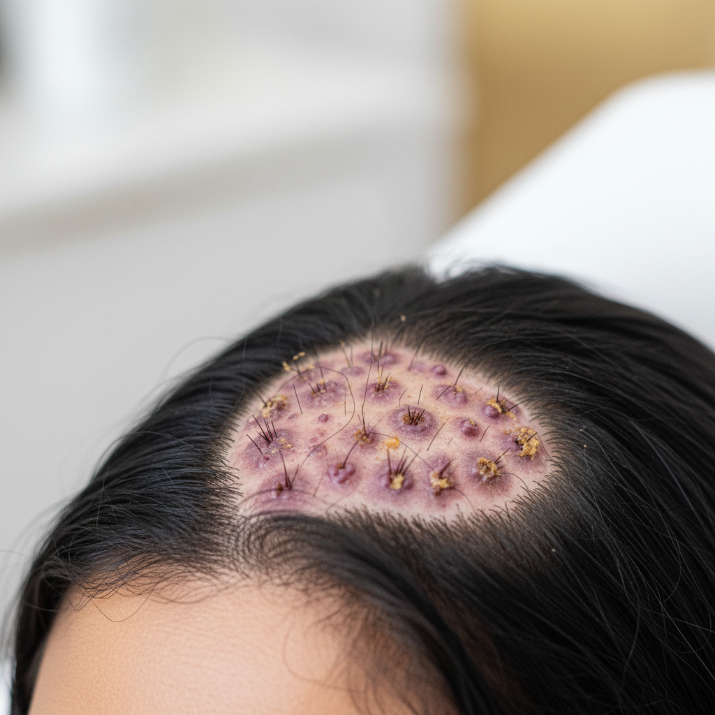
Early follicular pustules -> Crusting and follicular keratosis -> Tufted folliculitis -> Fibrotic scar tissue
The clinical progression typically involves:
- Perifollicular erythema and pustules: Small, tender, yellowish pustules erupt around the hair bases, primarily at the crown, vertex, or occipital regions of the scalp.
- Crusting and bleeding: Pustules rupture easily, forming thick, adherent yellow or honey-coloured crusts. Removal of these crusts often provokes pinpoint bleeding.
- Pain, burning, and pruritus: Patients frequently report persistent throbbing, tenderness, or intense itching across the affected scalp zones.
- Tufting (Polytrichia): Multiple hair fibres emerge from a single follicular ostium. This phenomenon occurs when destruction of interfollicular dermis forces adjacent follicles to fuse into a single fibrous tract.
- Induration and scarring: The scalp becomes boggy or firm to the touch, eventually settling into smooth, ivory-white or pinkish atrophic plaques devoid of follicular pores.
How folliculitis decalvans compares to other scalp conditions
Because early symptoms mimic more benign scalp issues, misdiagnosis is common. The table below outlines distinguishing features across common inflammatory scalp presentations.
| Condition | Primary Mechanism | Scarring? | Hallmark Clinical Sign | First-Line Management |
|---|---|---|---|---|
| Folliculitis Decalvans | Neutrophilic scarring inflammation | Yes (Permanent) | Tufted hairs emerging from single pore; yellow crusts | Oral antibiotics (e.g. Clindamycin + Rifampicin) |
| Bacterial Folliculitis (Superficial) | Simple bacterial infection of ostium | No (Temporary) | Small, discrete pustules without hair loss | Topical antiseptics; short-course topical antibiotics |
| Dissecting Cellulitis | Follicular occlusion tetrad component | Yes (Permanent) | Interconnecting fluctuant nodules, scalp bogginess, sinus tracts | Oral isotretinoin; biologics; surgical excision |
| Lichen Planopilaris | Lymphocytic scarring inflammation | Yes (Permanent) | Perifollicular violet erythema, keratotic plugs, no pustules | Topical/intralesional steroids; hydroxychloroquine |
| Scalp Psoriasis | Hyperkeratosis / auto-inflammatory | No (Temporary shedding possible) | Thick, silvery-white plaques with well-demarcated borders | Topical corticosteroids; calcipotriol; coal tar |
Medical treatment and scalp management
Achieving remission requires prescription medical intervention managed by a dermatologist. The primary objective is to extinguish active neutrophilic inflammation and reduce S. Aureus bioburden.
Systemic pharmaceutical approaches
Peer-reviewed dermatological consensus strongly favours combination antibiotic therapy. The most well-documented regimen is oral rifampicin (300 mg twice daily) combined with oral clindamycin (300 mg twice daily) for a duration of 10 to 12 weeks. Rifampicin achieves high intracellular concentrations and clears staphylococcal carriage, while clindamycin contributes anti-inflammatory properties alongside its antimicrobial spectrum.
Other systemic options include:
- Tetracyclines (Doxycycline, Minocycline): Often deployed in milder or early-stage disease primarily for their matrix metalloproteinase-inhibiting and anti-inflammatory effects.
- Oral Isotretinoin: Prescribed in recalcitrant cases to alter the follicular microenvironment, reduce sebaceous gland volume, and induce long-term remission.
- Biologics and Dapsone: Used off-label for severe, refractory disease unresponsive to standard antimicrobial protocols.
Supportive scalp care and hygiene
While medical therapy does the heavy lifting, supporting the scalp barrier is crucial. Patients should use lukewarm water rather than hot water, which can trigger vasodilation and exacerbate scalp burning. Gentle, physiological-pH (pH 5.0 to 5.5) cleansers help remove secondary crusting without stripping the delicate epidermal barrier. Antiseptic washes containing chlorhexidine or diluted povidone-iodine may be recommended by clinicians to lower surface colonisation, but they must be balanced to avoid contact dermatitis.
Supporting hair and scalp health: The Watermans approach
When individuals experience chronic scalp disorders like folliculitis decalvans, the surrounding, unaffected follicles remain vulnerable to secondary stress. Physiological inflammation and psychological distress can precipitate telogen effluvium across unaffected zones of the scalp.
In such circumstances, maintaining the cleanest, most supportive environment for uninvolved hair is critical. Watermans focuses on providing nutrient-rich, scalp-first cosmetic formulations. The Watermans GrowMore Elixir and GrowMe Shampoo are engineered with ingredients such as biotin, rosemary, caffeine, and niacinamide.
While cosmetic products must never be viewed as a medical treatment or cure for folliculitis decalvans, maintaining optimal scalp hygiene, nourishing non-scarred follicles, and improving hair fibre condition supports a fuller-looking, healthier appearance alongside prescribed dermatological care.
When to seek immediate dermatological advice
If you or a salon client observe persistent follicular pustules accompanied by scalp soreness, crusting, or tufts of hair bunching together, do not adopt a "wait and see" approach.
Every week of active, uncontrolled pustular inflammation risks further destruction of irreversible stem cells within the hair follicle. Stylists should refrain from applying chemical services, bleach, permanent hair colour, or high-tension styling to inflamed zones. Instead, recommend an urgent evaluation by a dermatologist or a registered trichologist. A definitive diagnosis is typically confirmed using dermoscopy (trichoscopy) and a 4 mm punch biopsy taken from the active inflammatory margin of a lesion.
Frequently asked questions about Folliculitis Decalvans Overview
Can hair regrow once folliculitis decalvans is cured?
No, hair cannot regrow in areas where the disease has run its course. As detailed in this Folliculitis Decalvans Overview, the condition is a cicatricial alopecia. The inflammatory process destroys the follicular stem cell niche in the bulge, replacing the entire hair follicle with fibrous scar tissue. Hair can only be retained in areas where follicles have not yet suffered complete fibrotic obliteration.
Is folliculitis decalvans contagious?
Folliculitis decalvans is completely non-contagious. Even though the bacterium Staphylococcus aureus is commonly isolated from the lesions, the root pathology stems from an idiosyncratic host immune and inflammatory reaction to the bacteria, rather than a transmissible infection. You cannot catch it from salon equipment, family members, or direct contact.
How is folliculitis decalvans diagnosed?
Dermatologists diagnose folliculitis decalvans through clinical examination, trichoscopy, bacterial swab cultures, and a scalp biopsy. Trichoscopy typically demonstrates follicular tufting, crusting, and perifollicular erythema. A 4 mm punch biopsy of an active, untreated pustular border confirms the presence of deep neutrophilic inflammation and follicular destruction.
Can diet or lifestyle changes manage folliculitis decalvans?
There is no clinical evidence demonstrating that dietary changes, vitamin supplements, or lifestyle modifications alone can arrest folliculitis decalvans. While an anti-inflammatory lifestyle supports general wellbeing, this condition represents an aggressive, deep-seated dermatosis that requires targeted pharmaceutical intervention to prevent lifelong disfigurement and baldness.
Navigating Folliculitis Decalvans
Folliculitis decalvans is an aggressive, challenging scalp condition that demands immediate clinical respect. As outlined in this Folliculitis Decalvans Overview, distinguishing between superficial scalp irritations and true primary cicatricial alopecia is paramount. Because every inflammatory flare compromises irreplaceable follicular stem cells, prompt medical diagnosis, bacterial culture, and combination antibiotic therapy form the foundation of successful disease control. By combining evidence-based dermatological treatment with gentle, non-irritating cosmetic care for unaffected hair, individuals can halt the spread of scarring and protect their remaining hair.
Sources and references
- British Association of Dermatologists. Folliculitis Decalvans Clinical Guidelines. 2021.
- Powell, J. J., et al. "Folliculitis decalvans including tufted folliculitis: a study of 42 patients." British Journal of Dermatology, 1999.
- Rambhia, P. H., et al. "Updates in the Medical Management of Folliculitis Decalvans: A Systematic Review." Journal of Clinical and Aesthetic Dermatology, 2019.
- Otberg, N., et al. "Folliculitis decalvans." Dermatologic Therapy, 2008.
- National Institute for Health and Care Excellence (NICE). Alopecia: Cicatricial (Scarring) Management Summaries. 2022.
Find these ingredients in Watermans products
- Niacinamide: you will find this ingredient in our Hair Mask.