Cicatricial Alopecia Pathology: Mechanisms, Histology, and Diagnosis
10 min read ·
Explore cicatricial alopecia pathology, from stem cell destruction and histology to clinical diagnosis, biopsy markers, and evidence-based scalp management.
Quick answer: Cicatricial alopecia pathology refers to the microscopic cellular mechanisms that drive scarring hair loss, where targeted inflammation permanently destroys the hair follicle stem cell niche in the infundibulum and isthmus. In cicatricial alopecia pathology, destructive inflammatory infiltrates replace active pilosebaceous units with fibrous scar tissue, leading to irreversible loss of follicular openings. The primary clinical limitation is that once fibrotic tissue replaces the follicle, hair cannot regrow; early histological diagnosis via punch biopsy is essential to arrest disease progression before permanent bald patches expand.
Key takeaways
- Cicatricial alopecia represents a group of rare, inflammatory disorders causing permanent, irreversible follicular destruction.
- The hallmark mechanism involves immune-mediated destruction of epithelial stem cells located in the follicular bulge and the sebaceous gland.
- Pathologists classify primary scarring alopecias by the predominant inflammatory cell type: lymphocytic, neutrophilic, or mixed.
- A 4 mm scalp punch biopsy extending into the subcutaneous fat, processed with both vertical and horizontal sectioning, is the gold standard for definitive diagnosis.
- Cosmetic products and regular hair supplements cannot restore scarred follicles; medical management focuses strictly on halting inflammation to preserve surviving hair.
Cicatricial Alopecia Pathology: what the evidence shows
Cicatricial alopecia pathology centres on the premature and permanent destruction of the hair follicle's regenerative machinery. In a healthy scalp, follicles cycle continuously through anagen (growth), catagen (regression), and telogen (rest). This cyclical regeneration relies entirely on multipotent stem cells sequestered in the follicular bulge, situated near the insertion point of the arrector pili muscle, just below the sebaceous gland duct.
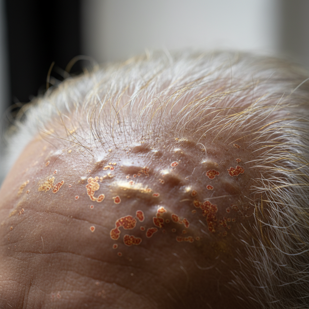
In primary cicatricial alopecia, an inflammatory cascade targets this specific anatomical zone. When immune cells invade the isthmus and infundibulum, the follicle collapses. The surrounding connective tissue sheath undergoes fibroplasia, filling the void left by the destroyed epithelial structure with dense collagen tracts. Under light microscopy, pathologists observe this end stage as fibrous tracts or follicular stelae devoid of hair shafts, internal root sheaths, or sebaceous glands.
Pathologists categorise primary scarring alopecia based on the North American Hair Research Society (NAHRS) consensus criteria, which splits conditions by the primary inflammatory infiltrate seen in active margins:
- Lymphocytic: Includes Lichen Planopilaris (LPP), Frontal Fibrosing Alopecia (FFA), Central Centrifugal Cicatricial Alopecia (CCCA), and Discoid Lupus Erythematosus (DLE). The infiltrate consists predominantly of CD4-positive and CD8-positive T lymphocytes attacking the follicular epithelium.
- Neutrophilic: Includes Folliculitis Decalvans and Dissecting Cellulitis of the Scalp (perifolliculitis capitis abscedens et suffodiens). Here, polymorphonuclear neutrophils predominate, frequently generating pustules, deep dermal abscesses, and sinus tracts.
- Mixed: Conditions such as Acne Keloidalis Nuchae and Acne Necrotica, displaying both lymphocytic and neutrophilic components alongside plasma cells and foreign-body giant cells reacting to free keratin from ruptured follicular walls.
What the evidence does not show
The pathology of scarring hair loss is often misunderstood in commercial salon settings. Scientific evidence does not support the idea that routine hair washing, cosmetic styling products, or typical hard water cause primary cicatricial alopecia. While mechanical traction can trigger traction alopecia, which may eventually scar if neglected for years, true primary cicatricial alopecias are intrinsic inflammatory autoimmune or auto-inflammatory disorders.
Furthermore, evidence does not show that hair loss in cicatricial conditions is reversible once the follicle has been replaced by fibrous tissue. No vitamin regimen, laser cap, or topical cosmetic formulation can regenerate an epithelial stem cell niche that has undergone complete fibroplasia. The clinical goal is preservation, not resurrection.
Cellular mechanisms of follicular destruction
To understand why cicatricial alopecia produces permanent baldness while conditions like alopecia areata allow for complete regrowth, one must examine immune privilege and stem cell biology.
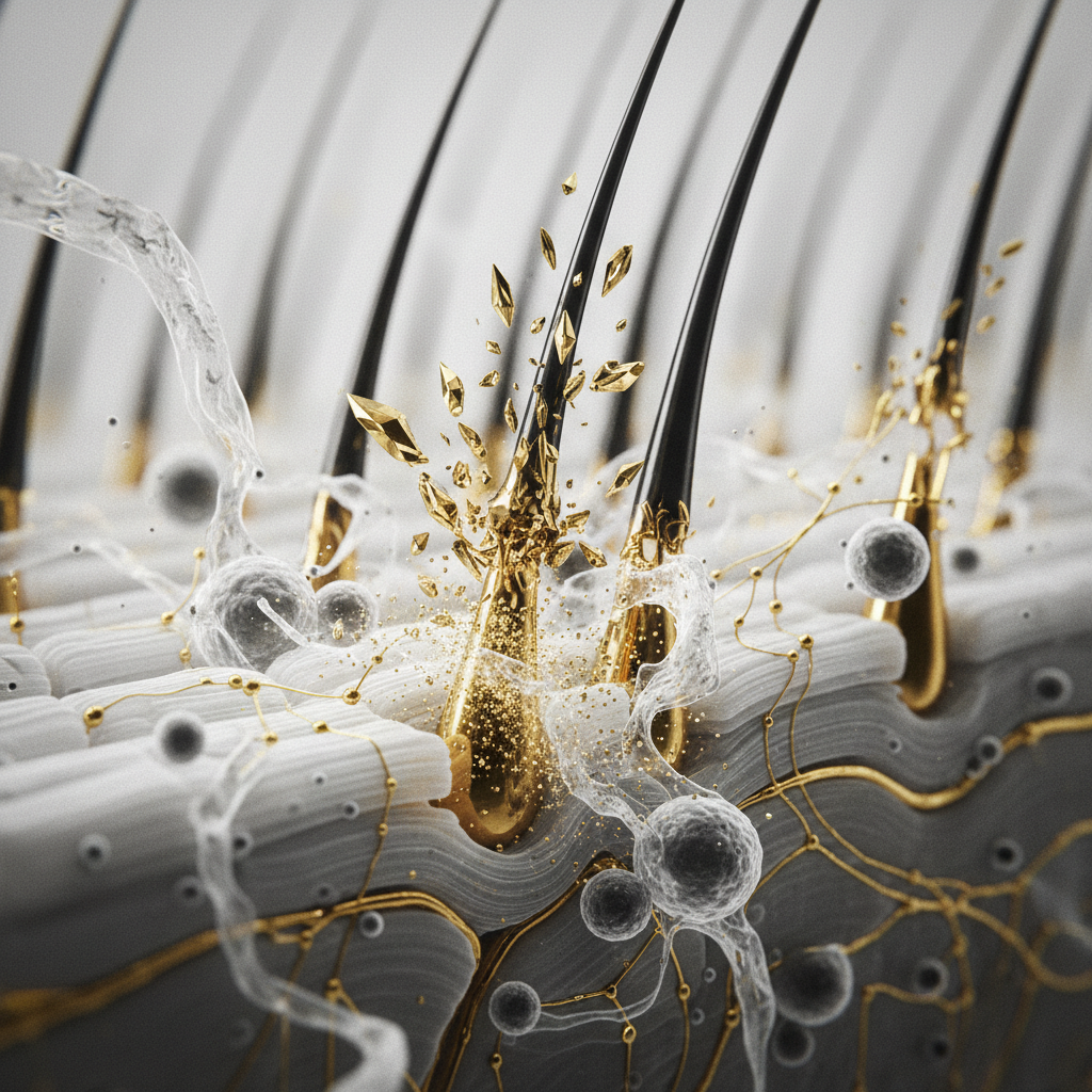
Healthy hair follicles maintain an immune-privileged status within the bulb region throughout anagen, down-regulating major histocompatibility complex (MHC) class I and class II molecules. In cicatricial alopecias like Lichen Planopilaris, this immune privilege collapses within the upper follicle. The follicular epithelium aberrantly expresses MHC class I and II antigens, prompting Langerhans cells to present follicular self-antigens to auto-reactive T lymphocytes.
As cytotoxic T cells accumulate at the junction between the follicular epithelium and the dermis, they induce apoptosis of follicular keratinocytes. Critically, this attack spares no mercy for the bulge stem cells expressing markers such as Keratin 15 (K15) and CD34. Simultaneously, the destruction of the adjacent sebaceous gland prevents the production of essential lubricating and barrier-protecting lipids. Peroxisome proliferator-activated receptor gamma (PPAR-gamma) expression, which normally maintains sebaceous gland homeostasis and lipid metabolism, drops sharply. Without functional sebaceous glands and bulge stem cells, the follicle loses all biological instructions for rebuilding an anagen hair shaft.
Fibroblasts migrate into the vacated dermal space, depositing thick bands of collagen types I and III. This process, known as cicatrisation, seals the follicular opening, causing the visible loss of follicular ostia on the scalp surface.
Histological patterns in scarring alopecias
The table below contrasts the classical microscopic and histological presentations seen across the primary forms of scarring alopecia.
| Condition | Primary Infiltrate | Anatomical Focus | Distinctive Microscopic Features |
|---|---|---|---|
| Lichen Planopilaris (LPP) | Lymphocytic (T cells) | Infundibulum and isthmus | Dense lichenoid interface dermatitis; apoptotic keratinocytes (Civatte bodies); concentric peri-infundibular lamellar fibroplasia. |
| Frontal Fibrosing Alopecia (FFA) | Lymphocytic (T cells) | Marginal frontotemporal hairline | Identical to LPP histologically; marked loss of sebaceous glands; apoptotic cells restricted to the upper follicle. |
| Discoid Lupus Erythematosus (DLE) | Lymphocytic (T and B cells) | Superficial and deep perivascular, periadnexal | Follicular plugging; basal layer vacuolation; thick periodic acid-Schiff (PAS) positive basement membrane; mucin deposition. |
| Central Centrifugal Cicatricial Alopecia (CCCA) | Lymphocytic | Middle to lower follicle, eccentric | Premature desquamation of the inner root sheath; eccentric thinning of the follicular epithelium; follicular rupture with giant cell response. |
| Folliculitis Decalvans | Neutrophilic | Upper follicle leading to deep dermis | Intrafollicular neutrophilic microabscesses; polytrichia (multiple hairs emerging from a single dilated ostium); dense plasma cell infiltrate later. |
Signs to look for: from dermoscopy to the clinic
Detecting cicatricial alopecia before irreversible destruction occurs requires a sharp eye for clinical and dermoscopic warning flags. Because the early stages mimic non-scarring telogen effluvium or seborrhoeic dermatitis, professional trichological assessment is crucial.
Key physical and trichoscopic indicators include:
- Peripilar casts: Tubular white or silver scales that wrap tightly around the base of the hair shaft (common in LPP and FFA).
- Perifollicular erythema: Redness directly encircling the follicular opening, pointing to active inflammation.
- Loss of follicular ostia: The scalp surface appears unnaturally smooth, shiny, or porcelain-like, with no visible pores.
- Polytrichia / Tufted folliculitis: Clusters of 5 to 20 hair shafts emerging from a single, dilated, scarred follicular pore, typical of advanced neutrophilic diseases.
- Scalp symptoms: Persistent burning, tenderness, pruritus, or a formication sensation (crawling skin) accompanying hair shedding.
What you can do and medical management
When managing scarring alopecia, time is tissue. The priority is rapid suppression of the destructive inflammatory infiltrate under the guidance of a dermatologist.
Medical treatments vary by primary cellular pathology:
- For lymphocytic forms (LPP, FFA, CCCA): High-potency topical corticosteroids (such as clobetasol propionate 0.05%), intralesional triamcinolone acetonide injections, oral hydroxychloroquine, or calcineurin inhibitors (tacrolimus) to calm cell-mediated auto-reactivity. PPAR-gamma agonists like pioglitazone may be considered to preserve sebaceous metabolism.
- For neutrophilic forms (Folliculitis Decalvans): Targeted oral antibiotic regimens (such as clindamycin paired with rifampicin) to eliminate underlying bacterial colonisation (often Staphylococcus aureus) and calm neutrophilic chemotaxis.
For cosmetic maintenance of surviving hair, gentle cleansing routines are recommended. Shampoos formulated without harsh sulfates, artificial fragrances, or high-pH detergents minimise barrier disruption on an already compromised scalp. Supporting the microcirculation of surviving, unaffected follicles can help maximise the density and visual presence of the remaining hair fibre.
When the cause may be something else
Not all patches of missing hair represent cicatricial alopecia pathology. Distinguishing between scarring and non-scarring hair loss is the most crucial step in differential diagnosis:
- Alopecia Areata: Characterised by sudden patchiness, but follicular ostia remain intact and open. Dermoscopy reveals yellow dots, black dots, and exclamation mark hairs. The follicle is miniaturised and held in an arrest phase, not destroyed.
- Androgenetic Alopecia: Shows progressive miniaturisation with follicular diameter diversity, without scarring, skin atrophy, or disappearance of follicular pores.
- Trichotillomania: Displays fractured hairs of varying lengths and normal scalp architecture under magnification, with no loss of follicular units.
- Tinea Capitis: Fungal infection causing hair breakage close to the scalp, marked by distinct fungal hyphae on potassium hydroxide (KOH) examination or fungal culture, though severe kerions can occasionally lead to secondary scarring if untreated.
When to get professional advice
If you or a client experience sudden or progressive patchy hair loss accompanied by scalp burning, soreness, or visible smoothing of the skin where pores are no longer detectable, seek an immediate referral to a consultant dermatologist.
The definitive diagnostic tool is a scalp biopsy. Two 4 mm punch biopsy specimens taken from the active, inflamed margin of an expanding lesion provide the tissue needed for comprehensive assessment. One sample is sectioned horizontally (transversely) to count follicular units and assess follicular density, while the second is sectioned vertically (longitudinally) to evaluate the dermo-epidermal junction and depth of inflammation.
The role of cosmetic hair care: Watermans support
While pharmaceutical intervention is the only method to halt the immune attack in cicatricial alopecia, cosmetic haircare remains important for protecting surviving strands and maintaining a calm scalp barrier.
Watermans products, such as GrowMe Shampoo, are designed as cosmetic scalp and hair formulations. Formulated with biotin, rosemary extract, niacinamide, and allantoin, these products do not treat or cure inflammatory medical alopecias. However, they provide a gentle, high-grade cleansing experience that cleanses sebum and debris without stripping the scalp. For individuals managing diagnosed scalp conditions alongside their medical therapies, maintaining scalp hygiene with non-irritating cosmetic cleansers supports overall fibre resilience and keeps non-involved follicles performing at their best.
Frequently asked questions about Cicatricial Alopecia Pathology
What is the primary difference in Cicatricial Alopecia Pathology compared to non-scarring hair loss?
The defining feature of cicatricial alopecia pathology is the irreversible immune-mediated destruction of epithelial stem cells in the hair follicle bulge, accompanied by destruction of the sebaceous gland and subsequent fibroplasia. In non-scarring alopecias, the follicle remains biologically intact and capable of regenerating new fibres once the triggering factor is resolved.
Can hair regrow once cicatricial alopecia scars the scalp?
No. Once cicatricial alopecia pathology results in complete replacement of the follicle by dense collagen fibrous tracts, the hair follicle is permanently destroyed. New hair cannot emerge from scarred dermal tissue. Medical treatment aims entirely to arrest inflammation and prevent active lesions from expanding into surrounding, healthy follicles.
Why is a scalp punch biopsy necessary to confirm the diagnosis?
A 4 mm punch biopsy is necessary because clinical examination alone cannot definitively separate lymphocytic from neutrophilic variants or identify early sub-clinical scar formation. Processing the biopsy with both horizontal and vertical sectioning allows a dermatopathologist to quantify follicular units, observe specific cell types, and pinpoint the anatomical level of destruction.
Does traction alopecia count as cicatricial alopecia?
Traction alopecia begins as a non-scarring mechanical condition caused by sustained tension on the root. However, if strong tension continues over many years, chronic inflammation and ischemic injury around the follicular bulb can eventually cause permanent scarring, transitioning the pathology into secondary cicatricial alopecia.
Overview of scarring hair loss
A comprehensive understanding of cicatricial alopecia pathology highlights the fragility of the follicular stem cell niche. Because inflammatory destruction of the isthmus and sebaceous gland leads to permanent fibrotic replacement of the follicle, early clinical identification remains the sole window of opportunity to preserve natural hair density. Combining prompt dermatological biopsy, targeted medical immunosuppression or antimicrobials, and non-irritating cosmetic maintenance provides the most reliable pathway to managing these challenging scalps.
Sources and references
- North American Hair Research Society (NAHRS). "Classification of primary cicatricial alopecias." Journal of the American Academy of Dermatology, 2001.
- British Association of Dermatologists (BAD). "Guidelines for the management of lichen planopilaris." British Journal of Dermatology, 2020.
- Harries, M. J., et al. "Management of primary cicatricial alopecias: options for treatment." British Journal of Dermatology, 2008.
- National Institute for Health and Care Excellence (NICE). "Alopecia - scarring and non-scarring presentations." Clinical Knowledge Summaries, 2021.
- PubMed Central. "Pathophysiology and histological evaluation of primary cicatricial alopecia." Dermatologic Clinics, 2013.
Find these ingredients in Watermans products
- Niacinamide: you will find this ingredient in our Hair Mask.