Alopecia Areata Diagnostic Markers: A Clinical Guide
7 min read ·
Understand how clinicians identify Alopecia Areata Diagnostic Markers using trichoscopy and biopsies, and how to spot key physical signs.

Quick answer: Clinicians identify Alopecia Areata Diagnostic Markers through a combination of physical signs, trichoscopic findings, and histological evaluations. Classic markers include exclamation mark hairs, yellow dots, and a peribulbar lymphocytic infiltrate resembling a "swarm of bees" on biopsy. While these indicators reliably confirm active follicular disruption, the primary limitation is that no single routine blood test can definitively diagnose alopecia areata or reliably forecast disease trajectory, meaning clinical and dermoscopic examination remain the diagnostic gold standard.
Key takeaways
- Alopecia areata is an autoimmune condition where T-lymphocytes target anagen hair follicles, leading to patchy, non-scarring hair loss.
- Dermoscopy (trichoscopy) provides rapid, non-invasive diagnostic markers including exclamation mark hairs, black dots, and yellow dots.
- Scalp biopsy remains the definitive tool for atypical presentations, showing a distinctive peribulbar lymphocytic infiltrate around growing follicles.
- Routine blood markers do not diagnose the condition, though doctors often test for comorbid autoimmune thyroiditis or coeliac disease.
- The presence of specific markers, like exclamation mark hairs, indicates active disease progression rather than chronic stability.
Alopecia Areata Diagnostic Markers: what the evidence shows
Clinicians rely on a layered approach to establish Alopecia Areata Diagnostic Markers, moving from gross clinical examination to magnified trichoscopy and, when necessary, histopathology. Alopecia areata occurs when the hair follicle loses its immune privilege. Cytotoxic CD8+ T cells cluster around the hair bulb during the anagen (growth) phase, driving the follicle prematurely into catagen and telogen phases. Because the follicle itself is not destroyed, this process is classified as non-scarring alopecia.
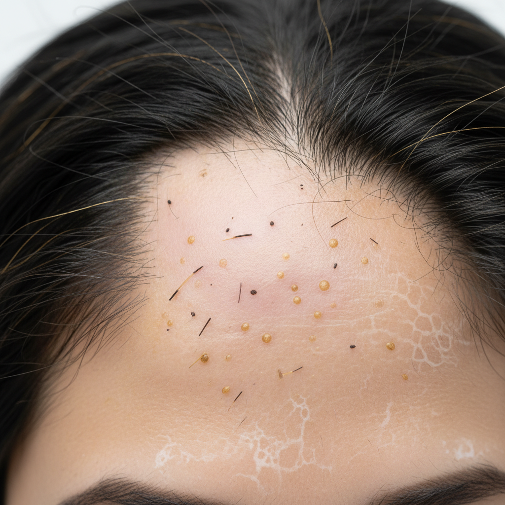
During physical inspection, doctors look for smooth, circular or oval patches of bare skin. Unlike fungal infections or scarring alopecias, the underlying skin typically shows no erythema, scaling, or scarring. A positive hair pull test at the margin of a patch suggests active disease, yielding telogen or dystrophic anagen hairs.
Under trichoscopy (magnification of 20x to 70x), several highly specific morphological markers emerge:
- Exclamation mark hairs (tapered hairs): These are short, fractured hairs (typically 1 to 4 mm long) where the proximal end near the scalp is thin and hypopigmented, while the distal tip remains thicker and darker. They represent a classic marker of acute, active disease.
- Black dots (cadaverised hairs): These appear when a hair fibre breaks at or just beneath the follicular opening due to rapid autoimmune damage before it can properly form.
- Yellow dots: These round, regular structures correspond to follicular infundibula distended with sebum and keratinous debris. They are visible in both active and chronic stages.
- Short vellus hairs: Thin, lightly pigmented regrowing hairs within the patch, indicating potential remission or early recovery.
- Broken hairs: Irregularly snapped shafts resulting from severe structural weakness caused by peribulbar inflammation.
When trichoscopy yields ambiguous findings, a 4 mm punch scalp biopsy offers definitive histological markers. Transverse sections reveal a dense lymphocytic infiltrate surrounding the lower pole of anagen and early catagen hair bulbs. Pathologists routinely describe this hallmark cellular arrangement as a "swarm of bees". Biopsies also display a dramatically shifted anagen-to-telogen ratio, with an elevated count of miniaturised and catagen follicles.
| Diagnostic Marker | Clinical Method | Disease Stage Indicated | Diagnostic Specificity |
|---|---|---|---|
| Exclamation mark hairs | Trichoscopy | Active / Rapidly progressing | High |
| Black dots | Trichoscopy | Highly active / Destructive | High |
| Yellow dots | Trichoscopy | Chronic or active | Moderate to High |
| Peribulbar lymphocytic infiltrate | Histopathology (Biopsy) | Active anagen disruption | Very High (Gold standard) |
| Short vellus hairs | Trichoscopy | Regrowth / Remission | Moderate |
| Dystrophic anagen roots | Hair pull test | Active margin spread | Moderate |
What the evidence does not show
The scientific literature does not support the idea that routine blood tests serve as primary Alopecia Areata Diagnostic Markers. While serum panels frequently check for ferritin, vitamin D, thyroid-stimulating hormone (TSH), and antithyroid peroxidase (TPO) antibodies, these do not diagnose alopecia areata itself. Instead, they identify potential systemic triggers, nutritional deficiencies, or associated autoimmune disorders.
Furthermore, biomarkers do not yet exist to predict how an individual will respond to specific treatments or whether a single patch will progress to alopecia totalis (total scalp loss) or alopecia universalis (total body loss). Elevated inflammatory cytokines, such as interferon-gamma (IFN-γ) and interleukin-15 (IL-15), are elevated in laboratory research settings, but commercial clinical assays lack standardisation for everyday diagnostic use.
Differential diagnosis: when the cause is something else
Because circular bald patches can stem from multiple mechanisms, doctors evaluate diagnostic markers to rule out lookalike conditions:
- Trichotillomania: Characterised by irregular, non-circular patches containing broken hairs of varying lengths, flame hairs, and coiled hairs on trichoscopy, without peribulbar lymphocytic infiltrates.
- Tinea capitis: A fungal infection featuring comma hairs, corkscrew hairs, scaling, and erythema, confirmed via potassium hydroxide (KOH) fungal scraping or fungal culture.
- Telogen effluvium: Involves diffuse, uniform shedding across the entire scalp rather than well-demarcated patches, triggered by metabolic stress, illness, or hormonal shifts.
- Lichen planopilaris (LPP): A scarring alopecia marked by perifollicular erythema, tubular peripilar casts, and the permanent loss of follicular openings.
- Syphilitic alopecia: Often referred to as "moth-eaten" alopecia, featuring patchy loss alongside systemic symptoms, diagnosed via serological testing (RPR/VDRL and treponemal assays).
When to seek medical advice
Anyone who discovers sudden, smooth circular patches of shedding should consult a dermatologist or a registered trichologist. Rapid intervention is particularly important if you observe active markers like exclamation mark hairs or if the shedding spreads quickly across multiple areas.
Medical evaluation is also essential if you notice nail changes. Pitting, trachyonychia (rough, sandpaper-like nails), or red spotted lunulae appear in roughly 10 to 20 percent of individuals with alopecia areata and often signify a more extensive clinical course.
A medical specialist can carry out dermoscopy, order appropriate blood tests to check for concurrent autoimmune thyroid disease, and discuss medical interventions such as intralesional corticosteroid injections, topical immunotherapy, or systemic JAK inhibitors where clinically indicated.
Supporting the scalp environment: cosmetic care
While autoimmune conditions require medical oversight, maintaining an optimal scalp environment supports overall hair fibre strength and skin comfort. Cosmetic products do not cure alopecia areata or reverse autoimmune follicle suppression. However, gentle, high-quality cleansing products can remove sebum buildup from follicular openings and prevent secondary microbial irritation.
Cosmetic formulations enriched with caffeine, biotin, niacinamide, and gentle botanical extracts can help nourish the surface of the scalp and condition existing strands. For example, Watermans Grow Me shampoo provides a revitalising, sulphate-free wash that respects the scalp's delicate barrier while improving the appearance of hair thickness and fibre resilience. Keeping non-affected hair strong and the scalp clean ensures that when medical remission occurs, regrowing fibres emerge into a balanced, healthy topical environment.
Frequently asked questions about Alopecia Areata Diagnostic Markers
What are the most common Alopecia Areata Diagnostic Markers seen under a dermatoscope?
The most reliable dermoscopic markers are exclamation mark hairs, black dots, and yellow dots. Exclamation mark hairs taper near the scalp surface and indicate active disease. Black dots represent hair fibres that snapped at the follicular orifice, while yellow dots reflect follicular openings filled with sebum and keratinous debris.
Can a regular blood test act as a diagnostic marker for alopecia areata?
No, standard blood tests cannot diagnose alopecia areata. Doctors use blood tests primarily to rule out co-occurring autoimmune conditions, such as Hashimoto's thyroiditis, or to identify underlying deficiencies in iron and vitamin D. Diagnosis relies on clinical observation, trichoscopy, and occasionally a scalp biopsy.
What is the histological gold standard among Alopecia Areata Diagnostic Markers?
The histological gold standard is a dense peribulbar lymphocytic infiltrate surrounding anagen hair bulbs, frequently described as a "swarm of bees". Pathologists identify this marker through a 4 mm horizontal punch biopsy of the scalp, which also reveals an abnormal ratio of catagen and telogen follicles compared to healthy tissue.
Do exclamation mark hairs mean my hair loss is getting worse?
Yes, exclamation mark hairs typically signal active, expanding disease. When clinicians identify these tapered structures at the outer margins of a bald patch, it indicates that the immune attack on nearby follicles is ongoing, suggesting the patch may continue to widen before stabilising.
How do doctors distinguish alopecia areata markers from androgenetic alopecia?
Androgenetic alopecia is distinguished by progressive follicular miniaturisation, resulting in hair diameter diversity greater than 20 percent across the crown or hairline. In contrast, alopecia areata markers feature distinct patches, yellow dots, black dots, and exclamation mark hairs without gradual pattern-based diameter variation.
Summary of Alopecia Areata Diagnostic Markers
Accurate identification of Alopecia Areata Diagnostic Markers is vital for distinguishing this reversible, autoimmune condition from irreversible scarring alopecias and fungal infections. Through dermoscopic signs like exclamation mark hairs and histological markers like peribulbar lymphocytic infiltration, clinicians can swiftly confirm a diagnosis and initiate appropriate therapy. While topical cosmetic care and supportive shampoos support external hair health, suspected cases of autoimmune hair loss should always be evaluated by a healthcare professional.
Sources and references
- British Association of Dermatologists. Guidelines for the management of alopecia areata. 2021.
- National Institute for Health and Care Excellence (NICE). Alopecia areata: Clinical knowledge summary. 2022.
- Rudnicka, L., et al. Atlas of Trichoscopy: Dermoscopy in Hair and Scalp Disease. Springer, 2012.
- Pratt, C. H., et al. Alopecia areata. Nature Reviews Disease Primers, 2017.
- American Academy of Dermatology. Alopecia areata: Diagnosis and treatment guidelines. 2020.
Find these ingredients in Watermans products
- Vitamin D: you will find this ingredient in our Hair Vitamins.
- Niacinamide: you will find this ingredient in our Hair Mask.