Understanding Lichen Planopilaris: Causes, Signs, and Management
9 min read ·
Understanding Lichen Planopilaris: discover the science, clinical signs, and medical treatments for this permanent, inflammatory scarring hair loss condition.
Quick answer: Understanding Lichen Planopilaris requires recognising it as a rare, inflammatory primary cicatricial (scarring) alopecia where the immune system attacks hair follicles. In this condition, cytotoxic T-cells destroy the epithelial stem cells located in the follicular bulge. Once these stem cells are replaced by fibrous scar tissue, the hair loss becomes permanent. The main limitation is that medical therapies cannot regenerate lost follicles. Early dermatological intervention aims solely to halt ongoing inflammation, ease painful symptoms, and preserve remaining hair.
Key takeaways
- Lichen planopilaris (LPP) is an autoimmune-mediated primary scarring alopecia that causes irreversible follicular destruction.
- It primarily affects adults between 40 and 60 years old, occurring more frequently in women than men.
- The condition specifically targets the follicular bulge, where epithelial stem cells reside, replacing active follicles with fibrous scar tissue.
- Common clinical signs include perifollicular erythema (redness), hyperkeratosis (scaling collars around hairs), burning, tenderness, and smooth patches without visible follicular openings.
- Cosmetic products and routine shampoos cannot cure or halt LPP; prompt clinical diagnosis by a dermatologist via dermoscopy and biopsy is vital to prevent ongoing permanent hair loss.
Understanding Lichen Planopilaris: what the evidence shows
Understanding Lichen Planopilaris begins with categorising it correctly within dermatology. LPP is considered the follicular variant of lichen planus, an inflammatory disorder affecting the skin, nails, and mucous membranes. In peer-reviewed literature, including guidelines from the British Association of Dermatologists (BAD), LPP is classified as a primary lymphocytic cicatricial alopecia.
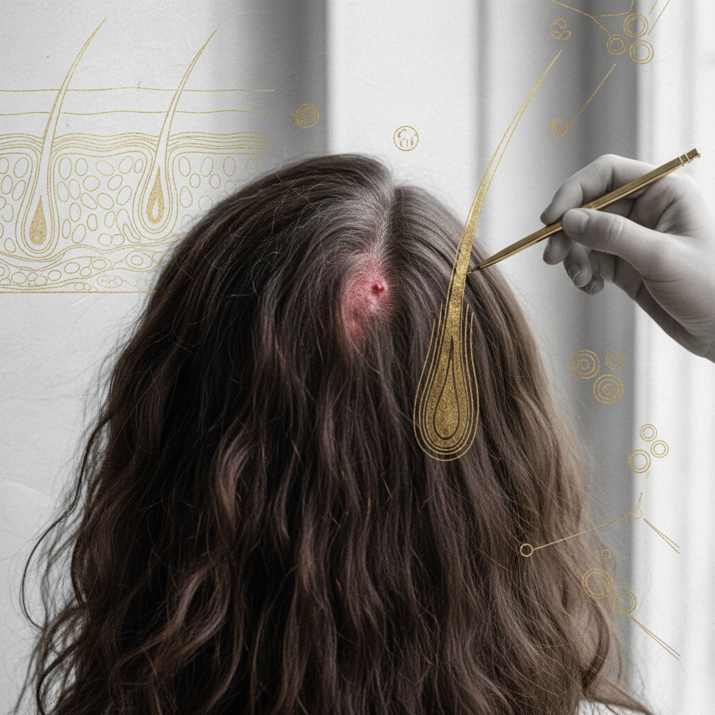
The pathological hallmark of this condition is a dense, lichenoid band of T-lymphocytic cells that infiltrates the upper part of the hair follicle. Dermatopathologists consistently observe that this inflammation zeroes in on the infundibulum and the isthmus of the follicle. Most critically, it targets the follicular bulge region. Because the bulge houses the multipotent epithelial stem cells responsible for cycling the hair follicle through anagen (growth), catagen (regression), and telogen (resting) phases, its destruction shuts down the regenerative capacity of the follicle forever.
Histological studies confirm that as these stem cells die, the body undergoes a fibroproliferative response. Collagen replaces the delicate architecture of the pilosebaceous unit. Under a dermatoscope (trichoscopy), this manifests as the total loss of follicular ostia (the tiny pores where hair emerges), leaving behind smooth, porcelain-white or ivory patches of scalp.
Three clinical variants of LPP exist in medical literature:
- Classic Lichen Planopilaris: Presents with patchy areas of scarring hair loss, predominantly on the vertex and crown of the scalp.
- Frontal Fibrosing Alopecia (FFA): Characterised by progressive, band-like recession of the frontal and temporal hairline, frequently accompanied by partial or complete loss of the eyebrows.
- Graham-Little-Piccardi-Lassueur Syndrome: A very rare triad involving cicatricial scalp alopecia, non-scarring pubic and axillary hair loss, and follicular keratotic papules on the trunk or limbs.
What the evidence does not show: myths and misconceptions
Much confusion exists surrounding scarring alopecias, leading individuals to mistake severe inflammation for common scalp conditions. The clinical evidence does not support several widespread assumptions:
- It is not standard dandruff or seborrhoeic dermatitis: While LPP involves scalp scaling, the scales form distinct, adherent collars around individual hair shafts (perifollicular casts), rather than broad, greasy flakes across the skin surface.
- It is not caused by poor hygiene or hair styling: LPP is an internal autoimmune reaction. It is not caused by infrequent washing, chemical dyes, heat styling, or hair care products.
- Cosmetics cannot regrow scarred follicles: No shampoo, serum, or botanical extract can resurrect a follicle once the stem cell niche has fibrosed. Over-the-counter hair growth products claiming to reverse scarring hair loss contradict clinical biology.
- It is not non-scarring alopecia: Unlike androgenetic alopecia (pattern hair loss) or telogen effluvium (temporary shedding), LPP destroys the follicular machinery entirely. Treating it like simple pattern loss delays life-saving interventions for the surrounding hair.
The biological mechanism: why the follicle scars
The destruction of the hair follicle in LPP stems from a collapse of follicular immune privilege. Healthy hair follicles enjoy an immunologically protected status, expressing very low levels of major histocompatibility complex (MHC) class I molecules. This protects them from auto-reactive immune surveillance.
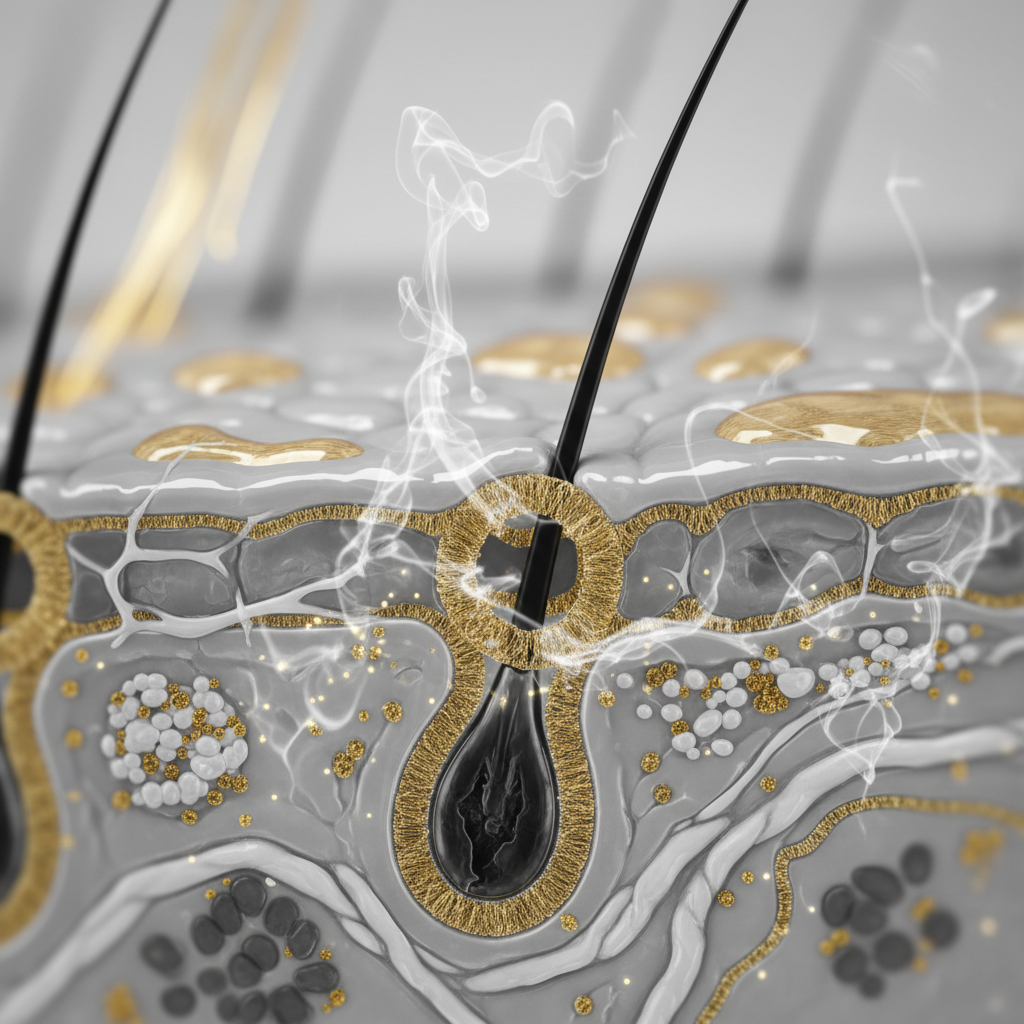
In LPP, this immune privilege breaks down. Studies show an abnormal upregulation of MHC class I and II antigens on the follicular epithelium. This alerts the immune system, drawing CD4+ and especially cytotoxic CD8+ T-lymphocytes directly to the outer root sheath.
These T-cells release pro-inflammatory cytokines, including interferon-gamma (IFN-γ), and trigger cell death pathways (apoptosis) in the follicular stem cells. As epithelial cells die, transformed fibroblasts synthesize dense type I and type III collagen. Eventually, the entire follicular tract is replaced by a fibrous tract (fibrous streamer), leaving an empty, scarred dermis where an active hair follicle once lived.
| Feature | Classic Lichen Planopilaris (LPP) | Androgenetic Alopecia (Pattern Loss) | Alopecia Areata |
|---|---|---|---|
| Alopecia Type | Primary cicatricial (scarring) | Non-scarring (miniaturisation) | Non-scarring (autoimmune shedding) |
| Primary Immune Target | Follicular bulge & stem cells | Androgen receptor pathways | Follicular bulb (hair matrix) |
| Follicular Openings | Absent in affected areas | Present (visible pores) | Present (visible pores, yellow dots) |
| Scalp Symptoms | Burning, pain, itching, redness | Usually asymptomatic | Mild itching occasionally |
| Hair Regrowth Potential | Zero once scarred | High (if miniaturised, not lost) | High (follicle remains intact) |
| Key Clinical Sign | Perifollicular erythema & casts | Miniaturised, wispy hairs | Exclamation mark hairs, smooth bald patches |
Signs to look for: clinical presentation
Identifying LPP early makes the difference between retaining a full head of hair and enduring widespread permanent baldness. Symptoms can develop slowly over years or escalate aggressively over weeks.
Key symptoms reported by patients include:
- Intense scalp tenderness or trichodynia (hair pain)
- Burning sensations that worsen when the scalp is touched or brushed
- Persistent itching (pruritus) that does not respond to regular anti-dandruff shampoos
Visible signs that a stylist or doctor might detect include:
- Perifollicular erythema: A rim of vivid pink, red, or violet inflammation encircling the base of individual hairs.
- Perifollicular hyperkeratosis: Hard, silvery-white keratin collars clinging tightly to hair shafts at the scalp surface.
- Loss of follicular ostia: Shiny, pale, smooth skin patches completely devoid of pore openings.
- Positive hair pull test: Gentle traction at the active margin of an inflamed patch easily dislodges anagen hairs with preserved root sheaths, signalling active destruction.
What you can do: clinical and supportive strategies
Because LPP is a serious medical disorder, primary care must always be directed by a medical professional. However, managing the condition requires a combined approach of medical therapy and gentle, supportive hair practices.
Medical interventions
Dermatologists aim to extinguish the fire of inflammation as quickly as possible. Evidence-based treatments documented in dermatological literature include:
- Topical and intralesional corticosteroids: High-potency topical steroids (such as clobetasol propionate 0.05%) or targeted triamcinolone acetonide injections directly into active margins to suppress local T-cells.
- Topical calcineurin inhibitors: Tacrolimus (0.1%) ointment to suppress T-cell activation without causing steroid-induced skin thinning.
- Systemic anti-inflammatories: Hydroxychloroquine (often prescribed at 200 to 400 mg daily), doxycycline, or short courses of oral corticosteroids to halt rapid progression.
- Immunosuppressants and modern targeted therapies: Mycophenolate mofetil, ciclosporin, or emerging Janus kinase (JAK) inhibitors for refractory cases under specialist hospital care.
Supportive daily care
While medical treatment addresses the immune attack, supportive routines help keep the surrounding scalp calm and prevent extra friction:
- Wash with mild, sulphate-free shampoos formulated around a physiological pH (roughly 5.0 to 5.5) to avoid irritating compromised skin barriers.
- Minimise thermal and mechanical stress. Avoid tight ponytails, harsh round-brush blow-drying, and heated styling tools close to inflamed patches.
- Avoid aggressive scalp scrubs or physical exfoliants. Vigorous friction can induce the Koebner phenomenon, where physical trauma triggers new autoimmune lesions in previously unaffected skin.
When the cause may be something else
Not all scalp redness or patchy shedding represents Lichen Planopilaris. Misdiagnosis is common, and differentiating LPP from mimics requires careful assessment:
- Discoid Lupus Erythematosus (DLE): Another scarring alopecia that features prominent follicular plugging, but often presents with deeper pigmentation changes, telangiectasia (spider veins), and scarring that extends into the interfollicular skin.
- Central Centrifugal Cicatricial Alopecia (CCCA): Scarring alopecia concentrated on the crown, seen predominantly in individuals of African ancestry, often driven by a distinct genetic predisposition and fibroproliferative pathway.
- Tinea Capitis: A fungal infection causing hair breakage, scaling, and inflammation that can resemble LPP, especially in children and young adults, easily confirmed or ruled out via mycology swabs.
- Folliculitis Decalvans: A neutrophil-driven scarring alopecia distinguished by painful pustules, yellow crusting, and tufted hairs (multiple hairs emerging from a single dilated pore).
When to seek professional advice
You should arrange an urgent appointment with a GP or consultant dermatologist if you notice shiny, hairless scalp patches, unexplained hair tenderness, or red rings around your hair follicles. Any sudden change in your frontal hairline or rapid loss of eyebrow density also warrants immediate specialist review.
Do not wait for hair shedding to worsen. In non-scarring hair loss, delaying treatment is unfortunate; in scarring conditions like LPP, delaying treatment means permanent follicular death. A dermatologist will perform trichoscopy and typically take a 4 mm punch biopsy from the active margin of the lesion to confirm the diagnosis histologically.
The role of cosmetic hair care
When managing Lichen Planopilaris alongside clinical treatment, keeping the surrounding, unaffected hair fibres strong and healthy remains important. While cosmetic products do not treat or prevent autoimmune scarring, they can reduce mechanical breakage along the remaining hair shafts.
Watermans focuses on gentle, non-irritating scalp care and strengthening treatments designed to support fuller-looking hair and maintain fibre resilience. Gentle cosmetic routines ensure that while medical treatments work to settle internal inflammation, the remaining hair is shielded from external damage, keeping the scalp environment clean and comfortable.
Understanding Lichen Planopilaris: ongoing management
Living with a chronic, cicatricial condition presents genuine emotional and physical challenges. Successfully Understanding Lichen Planopilaris involves recognising that although the disorder is chronic and irreversible in scarred areas, medical treatments can successfully push the condition into remission. Working closely with a qualified dermatologist, adopting protective daily styling habits, and monitoring active margins ensures you preserve the maximum amount of healthy, growing hair.
Frequently asked questions about Understanding Lichen Planopilaris
Can hair regrow once Lichen Planopilaris has caused a bald patch?
No. Once Lichen Planopilaris destroys the follicular bulge and replaces the follicle with scar tissue, regrowth is biologically impossible. The primary goal of medical management is to stop the inflammatory spread to save your remaining, healthy follicles.
How is Lichen Planopilaris definitively diagnosed?
A dermatologist confirms LPP through trichoscopy (evaluating follicular casts and erythema) followed by a scalp punch biopsy. A 4 mm skin biopsy taken from an active margin reveals the characteristic lichenoid lymphocyte infiltration around the infundibulum and bulge.
Does diet play a role in Understanding Lichen Planopilaris?
While anti-inflammatory diets support overall immune health, there is no clinical evidence that specific dietary adjustments can cure or reverse Lichen Planopilaris. Diet must never replace prescribed dermatological therapies such as corticosteroids or hydroxychloroquine.
Is Lichen Planopilaris contagious?
No, Lichen Planopilaris is entirely non-contagious. It is an autoimmune, inflammatory condition driven by your body's own immune cells attacking hair follicles. It cannot spread from person to person through touch, shared brushes, or salon tools.
Sources and references
- British Association of Dermatologists (BAD): Guidelines for the management of lichen planopilaris, 2020.
- National Health Service (NHS): Lichen planus and hair loss overview, 2022.
- Journal of the American Academy of Dermatology (JAAD): Cicatricial Alopecia: Clinicopathologic review and management guidelines, 2018.
- British Journal of Dermatology (BJD): The pathogenesis and clinical presentation of frontal fibrosing alopecia and lichen planopilaris, 2021.